The Fingerprint Band, Read by a Camera That Never Sees It
A chemical map without a dye
Infrared light between roughly 6 and 10 micrometres is absorbed by the specific chemical bonds that hold proteins, lipids and nucleic acids together, which makes the mid-infrared fingerprint band the part of the spectrum where biological material is most distinguishable without stains, antibodies or fluorescent labels. The reference protocol for the method describes it as a non-perturbative, label-free way to extract biochemical information aimed at diagnosis and at assessing how cells are functioning, and the sample survives the measurement. It has never become routine hospital equipment, and one important barrier is the detector rather than the chemistry.
The camera problem, answered sideways
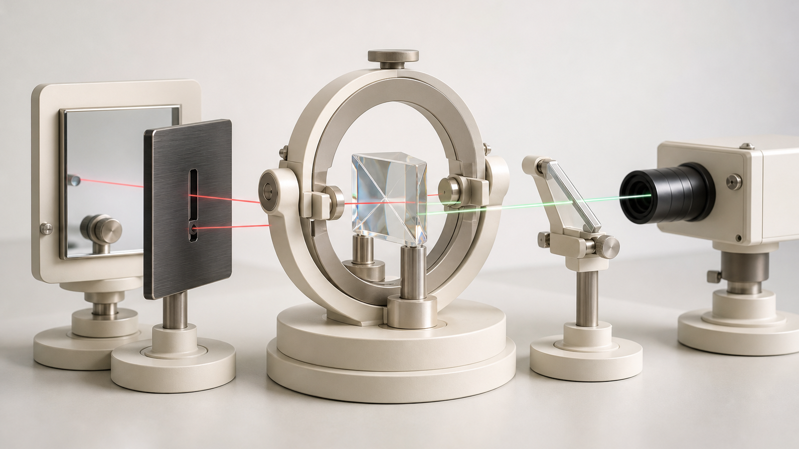
Detectors for that band are cooled, costly, and limited by the thermal glow of the room and of the instrument itself, because everything at ordinary temperature radiates in exactly the wavelengths being measured. A group at Imperial College London has now reported wide-field imaging across the full 6 to 10 micrometre range in which the infrared light is never measured at all. Correlated photon pairs are produced in a single silver thiogallate crystal used twice in a folded geometry, one partner passes through the sample, and the picture is reconstructed from the visible partners of those undetected photons on a commercial scientific silicon camera. Because the measurement happens in the visible, where the room's thermal background is effectively absent, the system detects infrared signals about a hundred times below the usual background-limited photodetection ceiling, at room temperature.
What the numbers actually allow
At 8 micrometres the images hold more than 8,000 resolvable elements at a resolution of 297 plus or minus 5 micrometres, over a circular field about 30 millimetres across, in a 10 second acquisition. The field is generous enough for a tissue section, a tablet or a culture well. The resolution is the constraint that matters: a human cell measures 10 to 20 micrometres, so that resolution is equivalent to roughly fifteen to thirty cell widths rather than to a camera pixel of that size, and the test objects were shadow masks cut from metal foil rather than biological material. This edition reads the result as a supply-chain and noise-floor contribution to an established clinical method, places it on the readiness ladder, and sets out the three specific demonstrations that would tell a hospital buyer the distance to a pathology bench is closing.