The Fingerprint Band, Read by a Camera That Never Sees It
Quentir Medicine Monitor
Evidence-based insights for quantum medicine. Published by Quentir Systems LLC · August 22, 2026.
A stain tells a pathologist where something sits in a tissue section. The mid-infrared fingerprint band tells you what that something is made of, and for decades the practical obstacle has been the camera rather than the chemistry.
Infrared light between roughly 6 and 10 micrometres is absorbed by the specific bonds that hold proteins, lipids and nucleic acids together. Reading that absorption gives a chemical map of a sample without dyes, antibodies or fluorescent labels. The detectors that see those wavelengths are another matter. They usually need cooling, they cost far more per pixel than a phone sensor, and they sit in the thermal glow of the room and of the instrument itself, because everything at ordinary temperature is radiating in exactly the band being measured.
A group at Imperial College London has now sidestepped that detector problem across the whole band. In a preprint posted on August 20, 2026, Vladimir Kornienko, Nathan Gemmell, Caiyi Liu, Asteria Chen, Chris Phillips and Rupert Oulton report wide-field imaging from 6 to 10 micrometres in which the infrared photons are never detected at all. The picture is reconstructed from the visible partners of those undetected photons, on a commercial scientific silicon camera.
Practical takeaway. The instrument reaches the chemical fingerprint band with a commercial silicon sensor in place of a cooled infrared array, and measures signals about a hundred times below the usual infrared background limit. Its resolution is 297 micrometres, which is fifteen to thirty times the width of a single cell, and the test objects were machined metal masks rather than tissue.
The band that carries the chemistry
Clinical infrared spectroscopy has a long and unglamorous history. The wavenumber range this work covers, 1670 down to 1000 reciprocal centimetres, is the part of the spectrum where biological molecules are most distinguishable from one another. The amide I and amide II bands near 1650 and 1550 reciprocal centimetres report on protein secondary structure. Ester carbonyl absorption reports on lipids. Phosphate stretching modes report on nucleic acids. A single spectrum from a spot of tissue therefore carries a compositional summary that no stain provides, which is why the method has been pursued in pathology research for decades.
The standard reference protocol for this kind of work, published by Matthew Baker and colleagues in Nature Protocols in 2014, describes the appeal in plain terms: infrared spectroscopy allows non-perturbative, label-free extraction of biochemical information from biological material, aimed at diagnosis and at assessing how cells are functioning. The sample survives the measurement, which is part of the attraction for tissue that is scarce or has to be kept for other tests.
One important barrier to routine hospital use is instrumentation. Mercury cadmium telluride focal plane arrays, the workhorse detectors of the band, need cryogenic or thermoelectric cooling, and their noise floor is set by the thermal photons streaming off the surroundings. That is the constraint the Imperial work attacks. It is not the only one, and the validation and workflow requirements discussed at the end of this piece would remain after any detector change.
Photography without detection
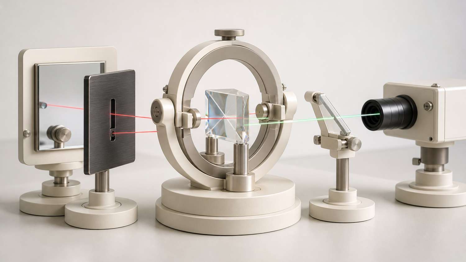
The method rests on an effect first demonstrated by Gabriela Lemos, Victoria Borish, Garrett Cole, Sven Ramelow, Radek Lapkiewicz and Anton Zeilinger, whose 2014 Nature paper on quantum imaging with undetected photons showed that an object can be imaged by light that is never measured. A nonlinear crystal produces correlated photon pairs. One partner is infrared and passes through the sample. The other partner is visible and goes to the camera. When the arrangement makes it fundamentally impossible to tell which of two possible emission events produced a given pair, those possibilities interfere, and the interference pattern recorded in the visible carries what happened to the infrared photon. The 2014 demonstration used two separate crystals for the two possibilities. The Imperial setup folds the same idea onto one. A single silver thiogallate crystal does the work twice, because a mirror sends all three beams back through it, so the two indistinguishable possibilities are the first pass and the return pass of the same crystal.
The technique has existed for a decade, mostly as narrow, point-by-point or small-field demonstrations. The step reported here is geometric. Non-collinear phase matching in the silver thiogallate crystal spreads the correlated pairs over angle rather than confining them to a single direction, and that turns a narrow probe into a wide field. The pump is a continuous-wave titanium-sapphire laser tuned between 700 and 740 nanometres at 50 milliwatts, and the detector is a Hamamatsu ORCA-Quest camera, a commercial scientific silicon sensor of the photon-number-resolving qCMOS class rather than a low-cost consumer part.
Quantum pillar: sensing. Technology readiness: TRL 4 of 9. The complete imaging system was assembled and tested on an optical bench against machined metal targets, with no biological sample, no patient material and no clinical setting involved so far.
A hundred times below the background limit
The performance claim worth understanding is the one about background. Conventional infrared photodetection has a ceiling called the background-limited infrared photodetection limit, which describes the point where the detector's sensitivity is capped by ambient thermal radiation rather than by the detector itself. The authors report imaging roughly a hundred times better than that limit, detecting infrared signals at least two orders of magnitude below it.
That number follows from the architecture. Because the infrared photon is never measured, the room's thermal photons in the 6 to 10 micrometre band never reach a detector that cares about them. The measurement happens in the visible, where the thermal background at room temperature is effectively zero, and the detection is coherent, so only light that interferes correctly contributes. The result is a room-temperature instrument reading a band that normally demands cooling.
Acquisition time separates two modes of operation. A single-frame holographic reconstruction took 10 seconds. A phase-scanning approach using 180 frames took about 30 minutes for a higher quality result. Both are laboratory numbers on a static target, and neither has been tested against a moving or degrading biological sample.
The number to keep is 297 micrometres
At 8 micrometres the images contain more than 8,000 resolvable elements, with a stated resolution of 297 plus or minus 5 micrometres, over a circular field of view of about 700 square millimetres, roughly 30 millimetres across. Those figures deserve to be read together rather than separately.
A field 30 millimetres wide is generous. It comfortably covers a standard tissue section, a tablet, a filter membrane or a culture well. The resolution is the constraint. A human cell is 10 to 20 micrometres across. Histopathology of the kind that changes a diagnosis works at micron scale or finer, because the diagnostic information sits in the arrangement of individual cells and nuclei. A 297 micrometre resolution is equivalent to roughly fifteen to thirty cell widths, which describes the finest detail the system can separate rather than the size of a camera pixel. What the instrument delivers today is a coarse chemical survey of a large area, well short of a cellular image.
There is a second honesty point in the paper that a press summary would drop. The test objects were shadow masks cut from 0.3 millimetre metal foil, chosen because they are binary transmitters with no phase shift, which makes the resolution measurement clean. No biological sample appears in this work. The authors are explicit that their result opens a path toward label-free biomedical imaging, materials science and chemical sensing. A path is what it is.
How Quentir Reads It
The interesting question is which axis actually moved. Infrared spectroscopic imaging of tissue is a mature research method with a thin clinical footprint, and its difficulty has never been the physics of absorption. It has been cost, cooling and thermal noise. This result does not improve the chemistry or the interpretation. It attacks the supply chain and the noise floor underneath them, which is a different kind of contribution and often a more durable one.
That distinction matters for anyone pricing the field. Two weeks of this Monitor have described quantum methods competing with established classical ones on accuracy, where the incumbent is strong and the margin is thin. Here the quantum effect changes which detector the band requires. Cooled focal plane arrays and up-conversion instruments already image these wavelengths, so the claim is narrower than a new capability: coherent detection in the visible, well below the thermal ceiling that limits direct photodetection, on a silicon sensor instead of a cooled infrared array. Neither the preprint nor the protocol supplies component or system pricing, and the bench described here still carries a tunable titanium-sapphire pump and specialized nonlinear optics, so no cost comparison is available yet. When a quantum technique substitutes a component rather than a judgment, cost and availability enter the argument early. Validation does not leave it. A replacement detector still has to prove analytical performance on real samples, fit an existing workflow, and satisfy whatever clinical evidence its intended use demands.
The humane stake is access rather than capability. Label-free infrared imaging of tissue has stayed largely inside research laboratories, and the cooling and cost of its detectors are among the reasons. An uncooled silicon route, if it survives the journey to micron resolution, would put chemical tissue assessment within reach of pathology services that are unlikely ever to buy a cryogenic focal plane array. Whether this route ends up cheaper is an open question the record does not yet answer, since the saving on the detector has to be weighed against a pump laser and nonlinear optics the conventional instrument does not need.
Three specific things would tell a hospital buyer that the distance is closing. Resolution has to improve by one to two orders of magnitude. The paper records that the 8,000 resolvable elements are equivalent to the number of modes in the down-conversion joint angular spectrum. That is an observation about where the mode budget sits, and the authors do not go on to show that the mode count is the operative limit, to rule out diffraction-related constraints, or to demonstrate that a different crystal and pump geometry would deliver the improvement a pathology bench needs. A real biological sample has to be imaged, with scattering, water absorption and thickness all present. And the spectral selectivity has to be demonstrated as a discrimination task, showing that two tissue states can be told apart, rather than as a tuning range. Until those exist, this belongs in the register as a strong physics result with a named clinical destination and no clinical evidence, which is the honest shape of most quantum medicine on the day it is published.
Sources
Primary source: Vladimir Kornienko, Nathan Gemmell, Caiyi Liu, Asteria Chen, Chris Phillips and Rupert Oulton, "Wide-field mid- to long-wave infrared imaging with undetected photons," arXiv preprint, submitted August 20, 2026, from Imperial College London. Method origin: Gabriela Lemos, Victoria Borish, Garrett Cole, Sven Ramelow, Radek Lapkiewicz and Anton Zeilinger in Nature, 2014. Clinical context for infrared analysis of biological material: Matthew Baker and colleagues in Nature Protocols, 2014.