A Weak Magnetic Field Left Its Mark Around a Tadpole's Eye
Quentir Medicine Monitor
Evidence-based insights for quantum medicine. Published by Quentir Systems LLC · July 24, 2026.
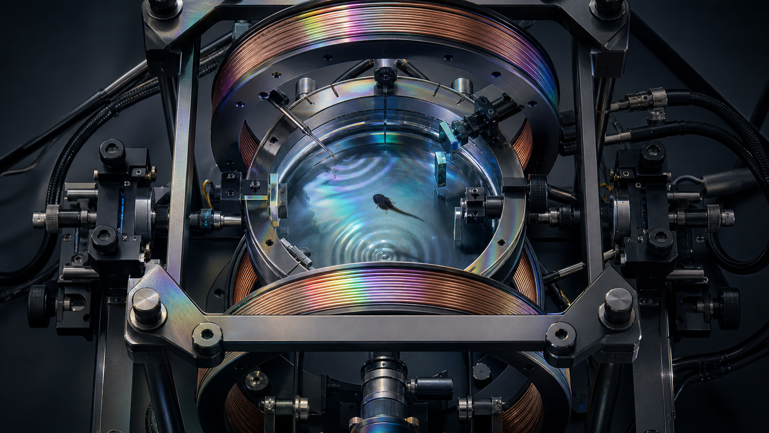
A tadpole raised on a white background changes its skin as its eyes read the light around it. In a Calgary laboratory, researchers placed that familiar developmental response inside a controlled magnetic field and counted what happened next.
The visible mark was a change in perioptic melanophores, the dark pigment cells around the eye. A July 16 bioRxiv preprint reports that fields between 0.25 and 1 millitesla increased those cells in a strength-dependent pattern. The light-and-eye dependence matters because it narrows the biology: the response vanished in constant darkness and after the eyes were removed. A spin-dynamics model could reproduce the curve, making a radical-pair mechanism feasible without identifying the molecule responsible.
Practical takeaway. This is a controlled whole-animal experiment in developmental biology, reported in a preprint. It offers a testable vertebrate model for quantum biology. It establishes no therapy, diagnostic, human-health effect, or clinical use.
A pigment cell becomes a biological meter
Xenopus laevis tadpoles have been laboratory workhorses for developmental biology because their embryos are accessible and their changing anatomy is easy to observe. Pigmentation is unusually useful here. Melanophores can move pigment within a cell over minutes, and the animal can also change how many melanophores develop over days. The new paper studies the slower, structural response around the eye.
Hadi Zadeh-Haghighi, Gabriel Bertolesi, Sarah McFarlane, and Christoph Simon raised embryos under a 12-hour light and 12-hour dark cycle, on a white background, inside or beside a Helmholtz-coil apparatus. The coils produced a uniform static magnetic field. The researchers kept field strength at or below 1 millitesla, controlled the temperature difference to less than 0.2 degrees Celsius, and scored images without knowing which treatment each tadpole had received.
At 0.5 millitesla, the number of perioptic melanophores rose by 37 percent across three independent experiments, with 28 to 34 tadpoles in each group. A separate experiment beginning earlier in development found a 75 percent increase at 0.75 millitesla under the light-dark cycle. The response appeared only after the retina had become functional. At 0.25 millitesla it was measurable; at 0.125 and 0.06 millitesla it was not statistically distinguishable from the controls at that time point.
Those values need context. The paper gives Calgary's local geomagnetic field as about 0.055 millitesla. The clearest pigmentation changes therefore appeared above the local background, and the authors state that their higher-field data do not solve the harder question of how a similar mechanism might work at geomagnetic strength. Field strength is part of the claim, not a footnote.
Light and the eye supply two useful controls
The strongest part of the experiment is the way its controls converge. Removing both eyes raised the baseline number of pigment cells by about 1.9 times, as previous developmental work would predict, and erased the additional magnetic-field response. Constant darkness also raised baseline pigmentation. In darkness, adding a 0.75-millitesla field produced no further significant increase.
The authors interpret that pattern as a shared pathway. Light received through the eye normally suppresses the development of these pigment cells. Darkness lifts the suppression. The magnetic field appears to lift part of the same suppression while light is present. The experiment leaves open where the field enters the pathway. Its controls make a nonspecific explanation such as simple heating less persuasive.
It also turns an elusive physical question into a countable biological outcome. A faint shift in chemical reaction yields would be hard to see directly inside a living tadpole. Researchers can count pigment cells blind and compare photographs across animals. That whole-animal readout is the paper's most useful contribution: it creates room for experiments that can break the proposed mechanism, not only fit it.
The quantum account fits, but does not identify the sensor
The proposed physics begins when light excites a flavoprotein such as cryptochrome. Electron transfer can create two short-lived radicals whose electron spins begin in a correlated state. Nearby atomic nuclei and an external magnetic field affect the balance between spin states. Because the spin state can influence which chemical products form, a weak field may alter the output of a biochemical reaction.
The authors fitted two models to four field points between 0.125 and 1 millitesla. One used a flavin-tryptophan radical pair parameterized from avian cryptochrome 4. The other used a generic organic radical pair. Both reproduced the rise and plateau in the pigmentation response. The generic pair fitted at least as well and needed much less biological amplification.
That ambiguity is important. The simulations show that radical-pair chemistry can produce the observed field-strength pattern with plausible parameters. They do not establish that cryptochrome 4 caused the pigmentation change. The paper says the link to CRY4 remains correlative until loss-of-function, radiofrequency-disruption, or wavelength-dependent experiments test it directly.
A peer-reviewed 2021 Nature study of European robin cryptochrome 4 supplies a related molecular anchor. That team found magnetically sensitive photochemistry in purified CRY4 in vitro, with stronger sensitivity than CRY4 from chicken or pigeon. The robin experiment validates a candidate spin-chemical system. The tadpole preprint adds a macroscopic vertebrate response. Neither result proves that frog pigmentation runs through the same protein.
Why this belongs near medicine, with a firm boundary
Quantum biology sits close enough to medicine to attract premature claims. Human tissues contain light-sensitive proteins and reactive molecules. They also carry electrical currents and trace magnetic materials. A credible effect in a vertebrate can therefore travel quickly from a developmental experiment to speculation about exposure, disease, or treatment.
The distinction between a biological response and harm is especially important here. The World Health Organization's electromagnetic-fields overview explains that a measurable biological response need not be an adverse health effect. The tadpole study counted a normal developmental cell type after controlled exposure. It reported no gross morphological difference after five days at 0.75 millitesla, and it did not study mammals, patients, disease, or long-term health.
Medicine may still gain something valuable from the model. Mechanisms that translate weak physical inputs into biochemical outputs are relevant to sensing, stimulation, and the design of experiments around light-sensitive proteins. A genetically tractable vertebrate system could help researchers ask whether the effect survives knockout of a candidate gene, changes with wavelength, reverses at predicted field strengths, or is disrupted by a resonant radiofrequency field.
Those are mechanism questions. Clinical relevance would need another chain of work: independent replication, molecular identification, dose and geometry mapping, tissue specificity, and a medically meaningful endpoint. The distance to care remains long, even when the underlying experiment is elegant.
How Quentir Reads It
Quentir reads the preprint as a falsification platform disguised as a pigmentation study. The authors have joined a controllable exposure, a blinded whole-animal readout, and a model that makes predictions beyond the fitted points. They predict a turnover and eventual sign reversal at higher fields, radiofrequency sensitivity, and a dependence on short-wavelength light if a flavin-based sensor is involved. A CRY4 knockout offers the cleanest route from correlation toward causality.
The lateral connection is to measurement science. Quantum medicine often starts with a device that detects a signal, as in Quentir's analysis of an engineered protein quantum sensor. Here the animal itself supplies the readout. That makes the setup less like a prototype instrument and more like an assay for deciding whether quantum spin chemistry survives the noise of a living vertebrate.
The next result that matters may be a failure. If the pigmentation response persists after CRY4 is disabled, ignores the predicted wavelength, or stays unchanged under radiofrequency disruption, the present explanation would weaken. If those tests behave as predicted and another laboratory reproduces them, the field would gain a stronger bridge between isolated-molecule spin chemistry and whole-organism biology. For now, a tadpole's dark cells have made that bridge visible enough to test.
Sources
Primary source: Zadeh-Haghighi, Bertolesi, McFarlane, and Simon, bioRxiv preprint posted July 16, 2026. Also drawn on: Xu and colleagues, Nature, June 23, 2021; and the World Health Organization electromagnetic-fields overview, accessed July 24, 2026.