Australia Funds a Quantum Window Into Living Brain Tissue
Quentir Medicine Monitor
Evidence-based insights for quantum medicine. Published by Quentir Systems LLC · August 10, 2026.
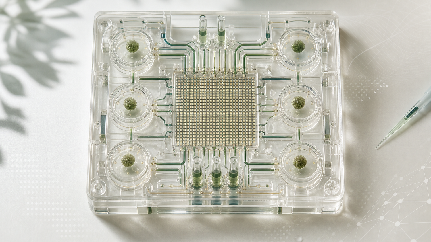
A neurological drug can look promising in a simplified cell assay and fail after it meets the electrical activity plus chemical and structural complexity of human neural tissue. The gap between a tidy laboratory result and a useful therapy is filled with models that reveal too little, too late.
An Australian consortium is building a brain-on-chip platform around that gap. The University of Melbourne and four companies plan to combine human neural micro-tissue with quantum tools that measure its activity in real time. The public description places the project squarely in preclinical drug discovery: a laboratory system for studying candidate therapies before anyone claims a clinical effect.
The idea is unusually concrete for quantum medicine. Living tissue supplies the biological model. A sensing layer follows electrical behavior. Drug developers gain a closer view of how neural tissue responds. The most valuable outcome would be a measurement system that catches a weak or harmful candidate before it reaches an expensive clinical program.
Practical takeaway. Australia has funded an integrated development project, not a treatment or diagnostic product. Its immediate test is whether the consortium can produce repeatable measurements from human neural micro-tissue that improve preclinical decisions.
The grant joins tissue engineering to a measurement problem
The University of Melbourne announced the consortium on March 3, 2026. The Australian government awarded it AUD 2.1 million through the Critical Technologies Challenge Program. University researchers are working with Chromos Labs, Tessara Therapeutics, Quantum Brilliance, and Axol Biosciences. The announcement names Alzheimer's disease, schizophrenia, epilepsy, and anxiety disorders as intended fields of use.
Each partner name points to a different part of the integration problem. The project needs living neural tissue that behaves consistently, hardware that can observe small signals, interfaces that preserve the tissue while measurements are made, and analysis that turns a dense time series into a useful preclinical judgment. A failure in any one layer can make the whole platform look noisier or more capable than it is.
A Team France Export account published on August 5, 2026 brought the project back into view and explicitly credits the University announcement. It is a derivative public account, not independent corroboration. The four named conditions differ sharply in biology and clinical course. Their presence should be read as the program's intended field of use, not as proof that one platform already models each disorder faithfully.
The model sits between a petri dish and a patient
A brain-on-chip is a laboratory model. Human neural cells or micro-tissues are arranged in a controlled device with channels, electrodes, or other sensing structures. A 2022 review in Biomaterials describes how such systems can model selected brain regions and the blood-brain barrier in normal or disease conditions. Researchers can expose the tissue to a candidate compound and watch what changes. The model retains more biological organization than a flat cell layer while remaining more controllable than a living organism. The review provides field context and does not establish the design or performance of this consortium's platform.
That middle position carries real value. Neurological drug development has to contend with human-specific biology, long disease courses, and endpoints that can be difficult to reproduce in animals. A micro-tissue model can narrow the distance between a molecular hypothesis and a human trial. It cannot reproduce a whole brain, a blood-brain barrier in all its complexity, a patient's immune system, or years of disease progression.
The consortium's public claim concerns real-time electrical activity in human neural tissue. That is a sensible target because neurons communicate through changing electrical states. A candidate therapy may alter firing patterns, coordination, or recovery after a controlled perturbation. Continuous measurement can show the timing and shape of a response that a single endpoint would miss.
Quantum pillar: sensing. Technology readiness: not applicable. The public record announces a funded development program and reports no integrated-system result, laboratory validation, or clinical study that can be placed on the readiness ladder.
The public record leaves the quantum interface underspecified
The announcement says that quantum technologies will support real-time measurement. It does not publish the sensor architecture, detection limits, sampling rate, noise floor, reference method, or the way the quantum component connects to the tissue model. Those omissions are ordinary at the opening of a funded project. They also define the technical questions that matter next.
Quantum Brilliance works across diamond quantum technology, but the cited project accounts do not identify a specific diamond defect, processor, or sensor configuration for this platform. A 2022 Nature Photonics paper by McCloskey and colleagues demonstrates the broader technical path: nitrogen-vacancy defects embedded in a transparent diamond device served as charge-sensitive fluorescent reporters for optical voltage imaging at biologically relevant voltages and timescales. That paper supplies technical background. It does not establish the consortium's final architecture. The defensible project classification is functional: the quantum contribution is presented as sensing because it measures a physical signal from tissue.
This distinction matters for hospital and pharmaceutical readers. A quantum sensor can be valuable without delivering a quantum-computing advantage. The project should eventually be judged against established measurement methods for the same tissue and task. Useful comparisons would include signal quality, stability over time, tissue viability, throughput and calibration burden, along with whether the measurement changes a drug-development decision.
The hardest engineering issue may be integration risk. A sensitive instrument can disturb the sample it observes. Microfluidic conditions can drift. Biological batches can vary. Electronics and analysis pipelines can turn ordinary noise into persuasive patterns. The consortium needs controls that separate a drug response from a sensor artifact, a tissue-preparation difference, or a software choice.
Why electrical activity matters to drug discovery
Many neurological therapies aim to change a system whose behavior unfolds over time. A compound may calm excessive firing, restore coordination, protect cells after stress, or produce an unwanted rhythm. Real-time recording lets researchers see direction, timing, and reversibility. That creates a richer picture than a final count of living cells.
Richer data can still mislead. A beautiful neural trace is only useful when it is reproducible and tied to a biological question. Researchers need blinded conditions, independent replication, known positive and negative controls, and a clear account of how raw signals become a claimed response. Disease-specific relevance matters too. An Alzheimer's micro-tissue model and an epilepsy model may require different cell compositions, perturbations, and endpoints.
For a drug developer, the commercial promise lies in earlier exclusion. Clinical trials consume years, money, and the time of patients and families who often have few options. A preclinical platform that rejects weak candidates sooner can redirect resources toward better ones. That humane benefit depends on accuracy. A false negative may discard a useful therapy, while a false positive carries a weak candidate further into animal work or human testing.
The program therefore has two customers before it has any patient. Scientists need a model they can trust, and development teams need a result that changes portfolio decisions. Hospitals may enter much later through trials, diagnostic research, or treatment programs. Keeping that clinical distance visible protects the project from the inflated language that often surrounds both brain technology and quantum technology.
How Quentir Reads It
Quentir reads this grant as an integration bet. Australia is placing public money where quantum sensing meets tissue engineering and drug-development economics. None of those domains carries the project alone. The tissue must model something biologically relevant. The instrument must measure it reliably. The resulting signal must improve a decision that conventional methods leave uncertain.
The original connection is between measurement and attrition. Drug discovery usually treats better measurement as a scientific benefit. Here it may also become an industrial filter. A sensitive, repeatable brain-on-chip platform could move failure earlier, when failure is cheaper and exposes no patient. That would be a meaningful contribution even if the platform never appears at a bedside.
The next credible milestone is modest and demanding: an integrated laboratory system, tested on human neural micro-tissue, with disclosed controls and comparison measurements. A disease claim should come later, after the model shows that it reproduces a relevant feature and that candidate compounds change that feature consistently. Clinical language belongs later still.
The grant opens a window onto living neural tissue. Whether the view is useful will depend on what the consortium can see repeatedly, what conventional instruments miss, and which development decisions change once the signal is available.
Sources
Primary source: University of Melbourne Newsroom, March 3, 2026. Derivative public account: Team France Export, August 5, 2026. Technical context: McCloskey et al., Nature Photonics, September 8, 2022; Amirifar et al., Biomaterials, April 21, 2022.