Siloton Announces £8.4 Million Innovate UK Contract on 14 October 2026 to Trial Its Chip-Based Handheld OCT Eye Scanner With Sunderland Eye Hospital
Siloton takes its chip-based retinal scanner toward NHS trials
Quentir Medicine Monitor
Evidence-based insights for quantum medicine.
The Bristol start-up Siloton has won an £8.4 million contract from Innovate UK to finish two versions of its handheld eye scanner, one for clinics and one for patients' homes, and to test both over the next two years. The company announced the award on 14 October 2026. The money comes through Contracts for Innovation: Quantum Missions Primer, a UK Research and Innovation program in which public bodies set a problem and pay companies to develop a solution.
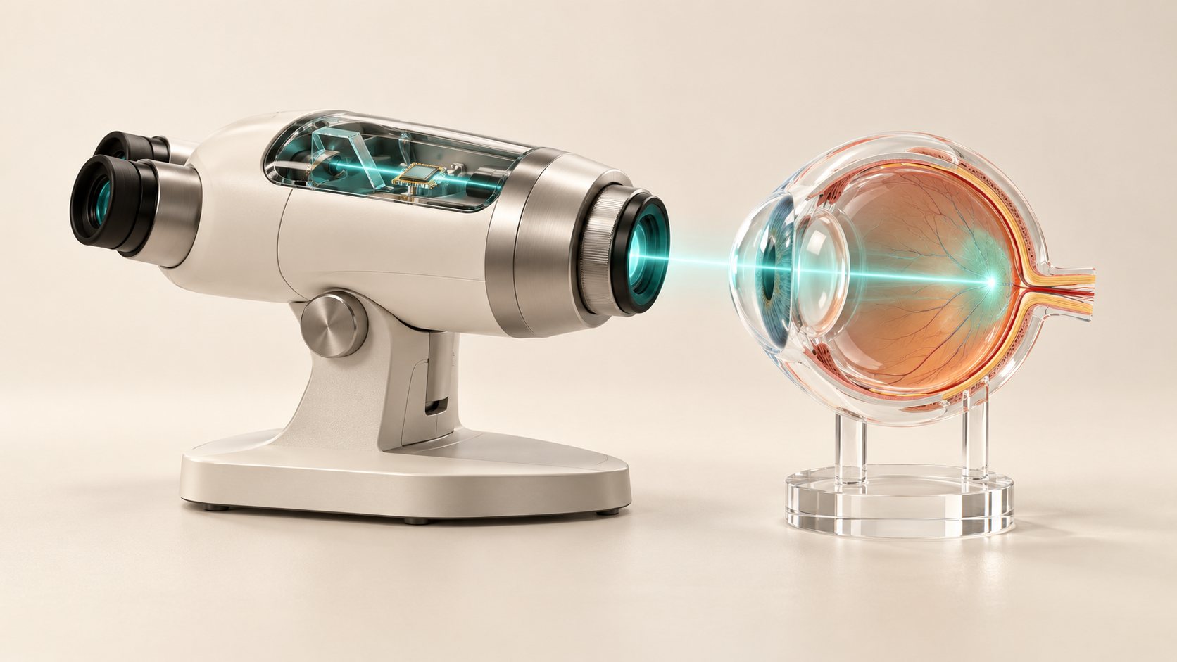
The device performs optical coherence tomography, or OCT, the scan that eye clinics use to see the layers of the retina in cross-section. Siloton says it has compressed a tabletop's worth of fragile optics onto a photonic chip smaller than a £1 coin, set in a binocular-style headset that a patient looks into. Clinical trials of the in-clinic version will run with Sunderland Specialist Eye Hospital, and York Health Economics Consortium will use the trial data to refine estimates of what the system could save the NHS.
For a hospital eye service, the question is practical. OCT is the main tool for following age-related macular degeneration, diabetic eye disease and glaucoma, and most patients on those pathways need repeat scans for years. A scanner small and cheap enough to sit in a community clinic, or in a living room, would change where those repeat visits happen.
According to Siloton's press release of 14 October 2026, the contract pays for the design of in-clinic and at-home versions of its GiraffeOCT device and for trials on both. Patients in the North East and North Cumbria will have access to the in-clinic system during the Sunderland trials. The company expects the funding to shorten its development by up to two years, and it plans to grow its staff to 16 by August 2028, with hiring in photonics, electronics and software.
Chief executive Alasdair Price said the contract also covers a wider range of detectable diseases, integration of a second-generation chip called BlueJay, which he described as the same architecture used in the company's system for astronauts, and a cloud platform behind the service. The European Space Agency is listed as a customer; it wants a way to follow the eye problems astronauts often develop in orbit. Siloton also plans a second-generation device, CheetahOCT, designed to measure further layers of the retina, and intends to raise more investment on the back of the award.
The company frames the need with NHS figures: ophthalmology is the busiest outpatient specialty in England, with about nine million attendances in 2023/24, and at the end of July 2026 nearly 623,000 people were waiting for an eye appointment, 8.7 percent of all NHS waits. These numbers come from the press release, which does not cite the underlying NHS dataset.
Zhejiang University Pairs Diamond Quantum Sensors With Structured Illumination to Map Oxidative Stress Inside Live HeLa Cells: Nature Communications, 10 October 2026
Diamond quantum sensors map oxidative stress inside live cells in Hangzhou
Quentir Medicine Monitor
Evidence-based insights for quantum medicine.
A team at Zhejiang University in Hangzhou has built a microscope that reads diamond quantum sensors inside living human cells at roughly twice the sharpness of conventional fluorescence imaging, and used it to watch reactive oxygen species change from one part of a cell to another. The paper, published in Nature Communications on 10 October 2026, reports that small diamonds in different regions of the same cell responded to two drug treatments in five distinct ways, a variety that a conventional averaged measurement smoothed into a single trend.
The performance figures in the abstract need careful reading, because they come from different experiments. The headline spatial resolution of 136 nanometers was measured on nanodiamond samples outside cells. Inside a live cell the team reports 161 nanometers for the optical image and 178 nanometers for the quantum sensing map. The 150 millisecond figure is the update interval of the sensing images, which the instrument reaches by reusing recent exposures in a rolling reconstruction. The 180 by 180 micrometer field of view is a mosaic of 5 by 5 smaller fields, captured in 12 seconds.
The article in Nature Communications (DOI 10.1038/s41467-026-78321-2) lists Yuxian Lu, Yibo Yang and Ke Xu as equal first authors, with Jiandong Feng as corresponding author. Most of the eleven authors work in the Laboratory of Experimental Physical Biology in Zhejiang University's Department of Chemistry, with co-authors from the university's College of Optical Science and Engineering, Zhejiang Normal University and China Jiliang University. The journal has released the accepted manuscript ahead of the final Version of Record.
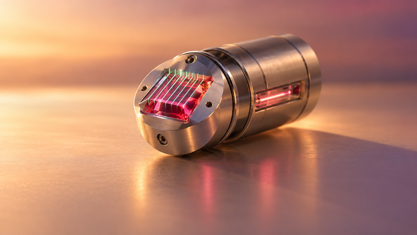
The sensing element is the nitrogen-vacancy center, a defect in diamond where a nitrogen atom sits beside a missing carbon atom. Its electron spin responds to temperature and to magnetic noise in its surroundings, and the defect reports that response as a change in red fluorescence. The team fed HeLa cells, a standard human cancer cell line, with fluorescent nanodiamonds about 100 nanometers across, which the cells took up over roughly a day.
IIT Bombay Review, 8 October 2026: Where Diamond Quantum Sensors and Fluorescent-Protein Qubits Stand for Medicine, From Living Cells to a Rat's Heartbeat
Quantum sensors for medicine, from diamond to fluorescent proteins
Quentir Medicine Monitor
Evidence-based insights for quantum medicine.
A cardiologist reads the heart through electrodes on the skin, and a neurologist reads the brain through sensors on the scalp. A small research field is trying to place quantum sensors next to the molecules themselves, inside cells and tissue, where they can report temperature, magnetic fields or chemical stress from within.
On 8 October 2026 three engineers at the Indian Institute of Technology Bombay posted a review of where that effort stands. Jyotiprakash Parhi, Ayan Majumder and Kasturi Saha follow quantum sensors for biomedicine from their established form, tiny defects in diamond, to a newer one: the fluorescent-protein qubit, a sensor a living cell can make for itself from its own genetic instructions. Between those two ends sit the nanodiamond probes that have already been placed inside cells to measure heat and the stiffness of the material around them.
The paper, "From Solid-State Spin Defects to Fluorescent Proteins: A Perspective on Quantum Optical Sensing Platforms for Biomedical Applications," is a preprint on arXiv (2610.11644) and has not been peer reviewed. It reports no new experiment. Its value for a hospital research director, a pathologist or a medical-device buyer is the map it draws: which measurements have been shown in cells, which in a living animal, and which problems the authors say still stand between these sensors and clinical use. A summary table sets out, sensor by sensor, the furthest biomedical stage reached, the representative study and the main barrier to translation.
The common thread is a defect or molecule whose electron spin changes how brightly it glows. Shine green or blue light on it, often together with a weak microwave signal, and the glow dims or brightens depending on the magnetic field, the temperature or the chemistry nearby. Because the readout is light, a microscope can collect it from inside a cell without wires.
QST's Heat-Etched Nanodiamond Quantum Sensors Cut Particle-to-Particle Variation to a Quarter and Raise the Signal 55 Percent: ACS Nanoscience Au, 7 October 2026
Uniform nanodiamond quantum sensors for blood diagnostics
Quentir Medicine Monitor
Evidence-based insights for quantum medicine.
A diagnostic test is only useful when it gives the same answer for the same sample, whichever batch of reagent the laboratory happens to open that morning. Tiny diamond particles that act as quantum sensors have shown remarkable sensitivity in research settings, yet each particle has behaved slightly differently, and that spread has stood between the laboratory result and a test a hospital could rely on.
On 7 October 2026 Japan's National Institutes for Quantum Science and Technology (QST), with Chiba University, Institute of Science Tokyo, Kyoto University and the Chiba start-up Type-I Technologies, reported a new way to make nanodiamond quantum sensors that behave far more alike. According to the QST press release of 7 October 2026, the new process reduces particle-to-particle variation to about a quarter of that of conventional particles and raises the quantum signal by 55 percent. The groups see the work as groundwork for quantum liquid biopsy, the detection of very small amounts of disease-related molecules in blood, and for batch production of uniform sensors rather than hand-picking a few outstanding particles.
The paper, "Spin-Uniform Nanodiamond Quantum Sensors for Reproducible Intracellular Thermometry," by Chihiro Suzuki, Ryuji Igarashi and nine co-authors, appeared online in ACS Nanoscience Au (DOI 10.1021/acsnanoscienceau.6c00068) on the same day. For a laboratory director, a diagnostics buyer or a clinician following early cancer detection, it addresses the question that comes before any claim of sensitivity: whether two sensors given the same sample report the same value.
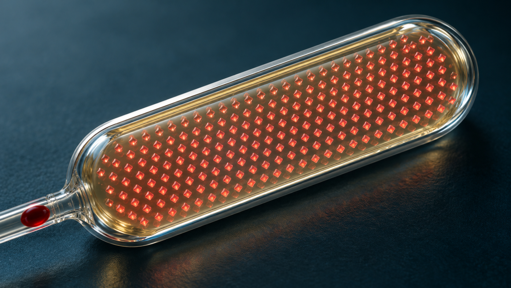
A nanodiamond sensor depends on a defect called the nitrogen-vacancy center, a spot in the diamond lattice where a nitrogen atom sits next to a missing carbon atom. When green laser light falls on the particle, the defect glows red, and microwave measurements of that glow, a method called optically detected magnetic resonance (ODMR), reveal small changes in temperature and in the chemical surroundings. Particles a few tens to a few hundred nanometers across can enter living cells.
The conventional route crushes larger diamond into nanoparticles first and creates the sensing defects afterward. QST explains that mechanical crushing leaves scratches and strain that differ from particle to particle, so every particle carries its own quirks into the measurement. The new process reverses the order. The team first builds nitrogen-vacancy centers into relatively large diamond particles of good crystal quality, then heats them gently in air so that the surface oxidizes away a little at a time. Diamond faces oxidize at different rates, and the researchers tuned the conditions so that disordered outer material goes first. What remains is the most orderly central tenth of the original particle, still small enough for work inside cells.
University of South Africa Quantum Dot Sensor Recovers Added SARS-CoV-2 Spike Protein From Wastewater Samples: The June 2026 Discover Electrochemistry Paper
Quantum dot sensor for SARS-CoV-2 in wastewater
Quentir Medicine Monitor
Evidence-based insights for quantum medicine.
Wastewater can show that an infection is spreading through a community before clinical case counts catch up. Reading that signal usually means shipping samples to a laboratory with PCR equipment and trained staff, which many rural districts and smaller municipalities do not have.
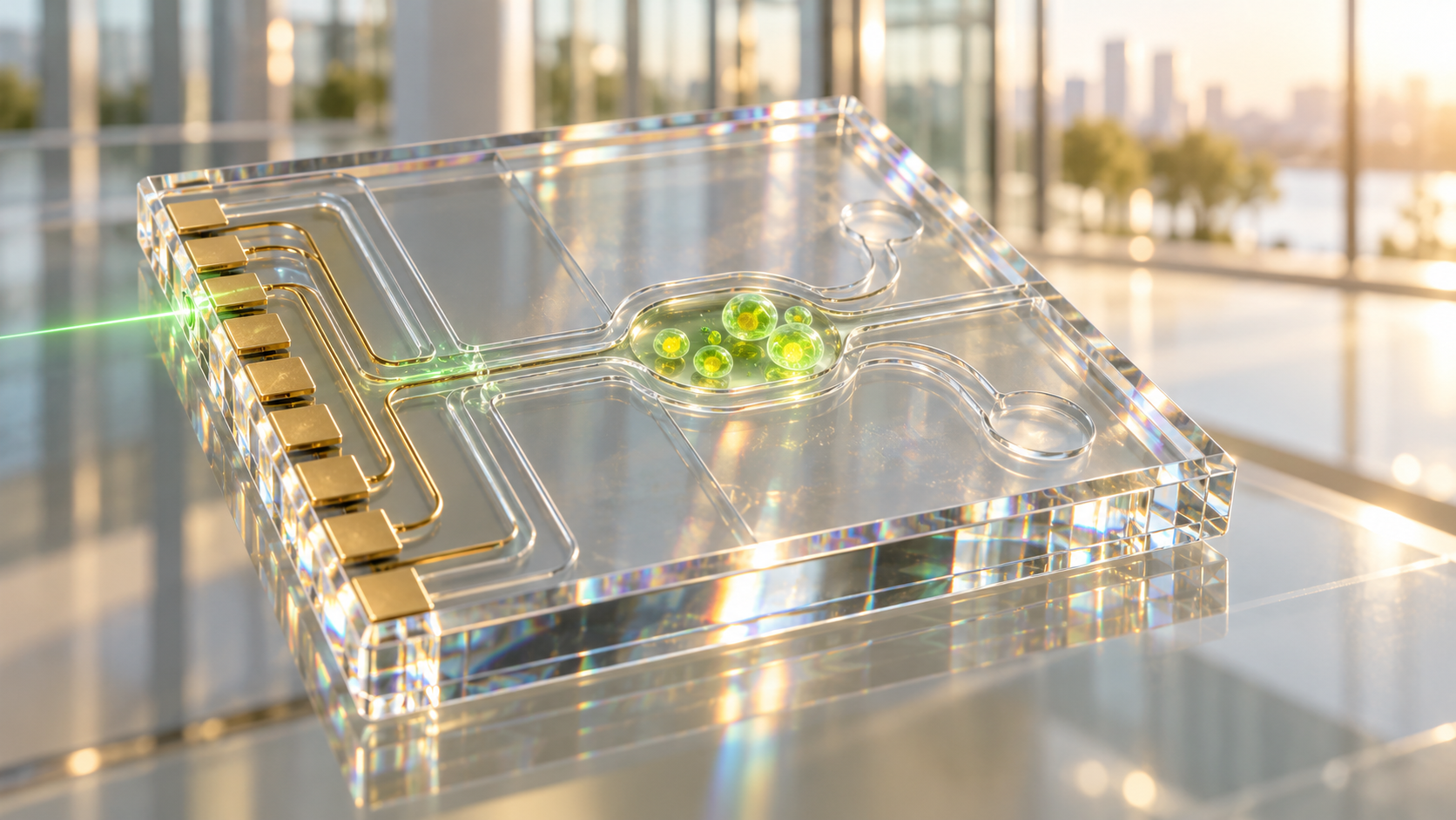
A team at the University of South Africa's Institute for Nanotechnology and Water Sustainability has built a small electrochemical sensor aimed at that gap. In a paper published in Discover Electrochemistry on 29 June 2026, Nolwazi T. Gazu, Xolile Fuku, Zikhona N. Cabunda, Bhekie B. Mamba and Usisipho Feleni describe an aptasensor that pairs the conducting polymer polyaniline with gadolinium telluride selenide quantum dots to detect the SARS-CoV-2 spike protein. The team reports a limit of detection of 0.04 femtomolar, measured in buffered saline, a figure that places the device among the most sensitive electrochemical approaches published for this target, according to a summary of the work that Bioengineer.org posted on 30 September 2026.
For hospital infection-control teams and public health laboratories, the interest lies in wastewater surveillance that could one day run closer to the treatment plant. The Monitor reads the paper as a careful laboratory result with a clear next step. The sensor has been tested on real effluent to which purified spike protein was added, and it now needs testing on wastewater that carries the virus naturally.
Polyaniline is a cheap, robust polymer that conducts electricity, which makes it attractive for low-cost electrodes. Its weakness is that conductivity drops sharply at neutral pH, the condition under which environmental water samples are usually analyzed. The researchers grew the polymer directly on a glassy carbon electrode and co-deposited the quantum dots with it. Quantum dots are semiconductor nanocrystals whose electronic properties follow from quantum confinement, the effect that appears when a crystal is only a few to a few dozen nanometers across. Here they act as electron mediators, lowering the resistance to charge transfer and compensating for the polymer's weakness at neutral pH. X-ray diffraction put the crystallite sizes between 10 and 32 nanometers, with an average of 17.

Kraków's J-PET Group and 21 Co-Authors Publish a Roadmap for Quantum Entanglement PET: What arXiv 2607.12182 and the June 2026 First-in-Human Scan Show
Quentir Medicine Monitor
Evidence-based insights for quantum medicine.
Every PET scan starts with a positron meeting an electron inside the patient. The two annihilate and send out a pair of gamma photons in opposite directions, and the scanner finds the tracer by catching both photons at once. Those photon pairs also carry a quantum property that no clinical scanner reads today: their polarizations are entangled. A group of physicists and imaging engineers now argues that this unused signal could sharpen PET images and add new diagnostic readings.
Their case is set out in "Roadmap Towards Quantum Entanglement Positron Emission Tomography (QE-PET)" (arXiv 2607.12182), posted on 13 July 2026 by 22 authors led by Paweł Moskal of the Jagiellonian University in Kraków, with teams from the University of California Santa Cruz, IIT Bombay, the University of Tokyo, the University of Sydney, the University of Zagreb, Japan's National Institutes for Quantum Science and Technology and others. The roadmap surveys how quantum entanglement PET could be built, from the plastic-scintillator J-PET scanner in Kraków to crystal, semiconductor and Compton-camera designs. It also describes a new candidate biomarker, the degree of quantum entanglement of the photon pair, which may depend on the tissue in which the positron annihilated.
Two experimental findings from the past three years made the idea worth pursuing. In 2023 an experiment showed that the quantum correlation between the two photons survives when one of them scatters off an electron, which most physicists had expected to destroy it. In 2025 the Kraków group reported in Science Advances that photons from annihilation in matter are less than maximally entangled, and that the degree of entanglement depends on how the annihilation happened. If that dependence tracks the chemistry of tissue, such as oxygen content, a scanner could read it as a new measurement alongside the usual tracer uptake.
A companion preprint makes the idea concrete. On 28 June 2026 the Kraków team and colleagues posted "First-in-human quantum entanglement imaging" (arXiv 2606.29421), reporting that the J-PET scanner produced, in one patient, both an image of a gallium-68 DOTA-TATE tracer and, from the same research acquisition, an image of the degree of entanglement. The roadmap reads that scan as a start. The authors state plainly that whether entanglement-based imaging and pH mapping can reach clinical practice "remains an open question."
Karystianou's Quantum Scanner Claim on Open TV, 20 September 2026: 100,000 Times Sharper Cancer Detection, Read Against the Mainz Diamond Magnetocardiogram
Quentir Medicine Monitor
Evidence-based insights for quantum medicine.
On 20 September 2026 Maria Karystianou, the pediatrician who leads the Greek movement Elpida gia ti Dimokratia, said on the Open TV program Tora Mazi, as transcribed by the newspapers Documento and Ta Nea, that the quantum scanners of the future will have 100,000 times greater resolution in locating cancer cells and will also be able to treat. She said it while the health minister, Adonis Georgiadis, was on the public broadcaster ERT calling her a "quantum doctor". The exchange is the newest turn in a Greek argument over the words quantum medicine that began with the death of the artist Giorgos Mazonakis and a private practice in Kolonaki, Athens.
Two parts of her statement are physics and hold up. The third is a forecast with no supporting instrument or publication identified in the sources reviewed here. This Monitor separates them. The quantum scanner claim of 100,000 times sharper cancer detection has no counterpart in the sources this Monitor reviewed; a quantum sensing result published on a human body this month, a diamond magnetometer reading of a heartbeat, is a separate research example that works at the scale of the whole heart and needs hundreds to thousands of beats to do it. What the episode shows a clinician is how the phrase quantum medicine now carries two meanings in one sentence.
The Greek material is unusually complete. Named physicists, a former university rector, a patients' union and a ministry have all spoken within one week, so a reader can see where the physics ends and the sales language begins. All quotations below are this Monitor's translations from the Greek newspaper accounts, which are secondary reports of the broadcasts; no broadcast recording was reviewed.
According to LiFO's account of the interview, which carries the physics passage and the inspection passage, Karystianou opened on inspections: after the Mazonakis case, she said, the state and its inspectors must check whether a practice is licensed and whether the procedures performed there are the ones a license covers, and the minister should stop telling patients to be careful about which practice they visit. Told on air that Georgiadis had just called her a "quantum doctor" on ERT, she answered that the word is quantum physics, that quantum physics has transformed science and technology, and that lasers are entirely quantum mechanics, as is the magnetic scanner, which she attributed to the quantum spin of protons.
Mainz, Stuttgart, Freiburg and Q.ANT Recorded a Human Magnetocardiogram With Room-Temperature Diamond Sensors: What the 16 September 2026 Science Advances Paper Shows
Quentir Medicine Monitor
Evidence-based insights for quantum medicine.
Three research groups in Germany have measured the magnetic field of a beating human heart with diamond sensing crystals smaller than half a cubic millimeter, at room temperature and without touching the skin. The paper appeared in Science Advances on 16 September 2026 under the coordination of Muhib Omar and Arne Wickenbrock of Johannes Gutenberg University Mainz, with Dmitry Budker and Jörg Wrachtrup among its 30 authors.
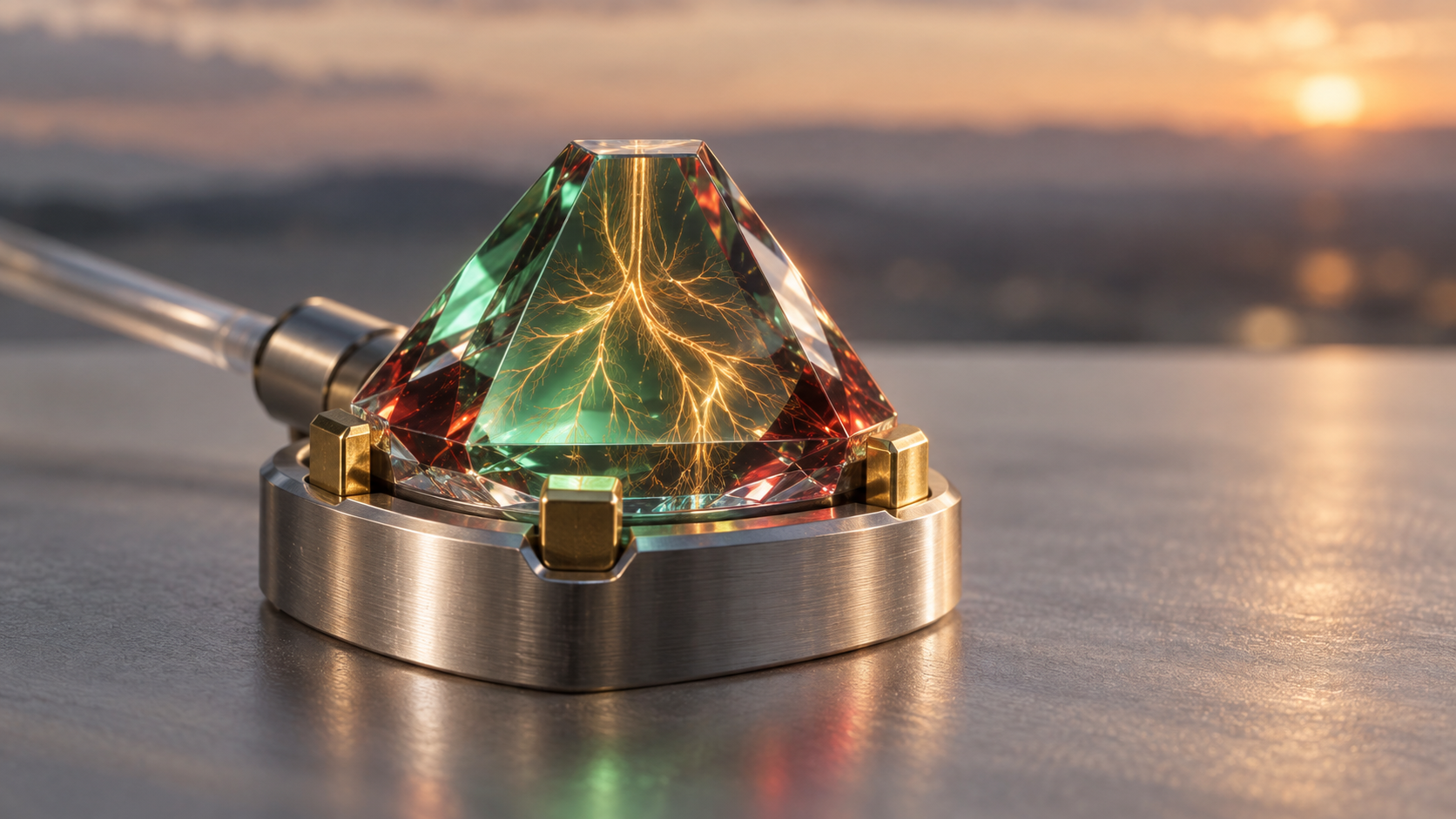
The instruments are diamond nitrogen-vacancy magnetometers: a nitrogen atom sitting beside a missing carbon atom in the diamond lattice, whose electron spin shifts its energy in proportion to the surrounding magnetic field. The three systems, built independently at Mainz, at the University of Stuttgart and by the Stuttgart startup Q.ANT GmbH, each produced a human magnetocardiogram in proof-of-concept self-experiments on members of the research groups, with sensitivities between 6 and 26 picotesla per square-root hertz. The result is a laboratory measurement on a handful of volunteers from the teams themselves, with no patient involved. It establishes that room-temperature diamond sensors can read the heart's field at all, after averaging, which is the step a cryogenic SQUID cleared decades ago and an optically pumped magnetometer clears with a heated vapor cell. The half-cubic-millimeter figure describes the sensing crystal, not the complete instrument with its fiber, optics and electronics, and the diamond heads recorded without touching the skin while a conventional electrocardiogram supplied the timing reference for the averaging, so the experiments did not show an electrode-free workflow.
The heart's magnetic signal is faint. The paper puts the average R-peak, the sharpest feature of each beat, at about 25 picotesla, roughly two million times weaker than the field of the Earth. That is why magnetocardiography has stayed inside hospitals that could afford superconducting detectors and the shielding around them, and why a SQUID magnetocardiograph still counts as deployable news when a Chinese manufacturer places one. The Mainz group's own line of work reached a related milestone on 6 August 2026 with a levitated-magnet sensor at 32 femtotesla per square-root hertz, a sensitivity reached only in a narrow resonance near 305 hertz, on a benchtop device that is far less suited than a diamond to sitting near a chest.
The Hellenic Physicists' Union Called "Quantum Medicine" an Unfounded Term on 12 September 2026: What the Word Still Covers, From Wearable MEG to Korea's Quantum Nanosensor
Quentir Medicine Monitor
Evidence-based insights for quantum medicine.
On 12 September 2026 the board of the Hellenic Physicists' Union adopted a press release titled "On the abuse of the term quantum" and posted it on its website the next evening. The one-page text lists what a quantum phenomenon is, credits the second quantum revolution with applications in biology and medicine, and then calls "quantum medicine", "quantum therapy" and "quantum consciousness" unfounded terms. It traces them to Deepak Chopra in 1989, cites Chopra's own 2007 statement that the phrase was a literary metaphor, recalls the 1998 Ig Nobel prize in physics, and adds that the degrees issued by a self-styled "Quantum University" in Hawaii are unrecognized, quoting McGill University's science office. Greece's health minister Adonis Georgiadis said the same on television on 15 September in plainer words: quantum medicine recognized by science does not exist.
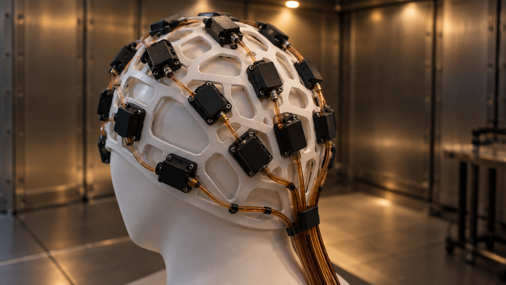
This Monitor publishes under the phrase the physicists reject, so the release deserves a careful reading, and a defensive one would miss what it concedes. The physicists are right about the word's history, and their release names no case, no clinic and no practitioner; Greek outlets reported it on 13 September alongside an Athens investigation, a connection the press drew and the release does not. The release also credits, in general terms, applications of quantum physics in biology and medicine. The Hellenic Physicists' Union statement separates a metaphor from measurable physics, and this Monitor applies that separation to two examples of its own choosing, which the union neither names nor endorses; that qualification is stated here once and holds for the whole piece. A wearable magnetoencephalography system built on optically pumped magnetometers, published in Nature in 2018, uses quantum sensing to measure brain magnetic fields and was demonstrated on healthy volunteers, with clinical use as the stated aim. A quantum nanosensor for cardiotoxicity funded by Korea's health ministry on 31 August 2026 is a funded research program with a named measurement still to be made, and no result yet. A practitioner's CV that lists a "quantum medicine" degree from an unaccredited online school is neither, and the difference is checkable by anyone who asks what was measured.
Hefei Mandi's Magnetocardiograph, 4 and 5 August 2026: Two Unresolved Deployment Descriptions in Chinanews and Xinhua
Quentir Medicine Monitor
Evidence-based insights for quantum medicine.
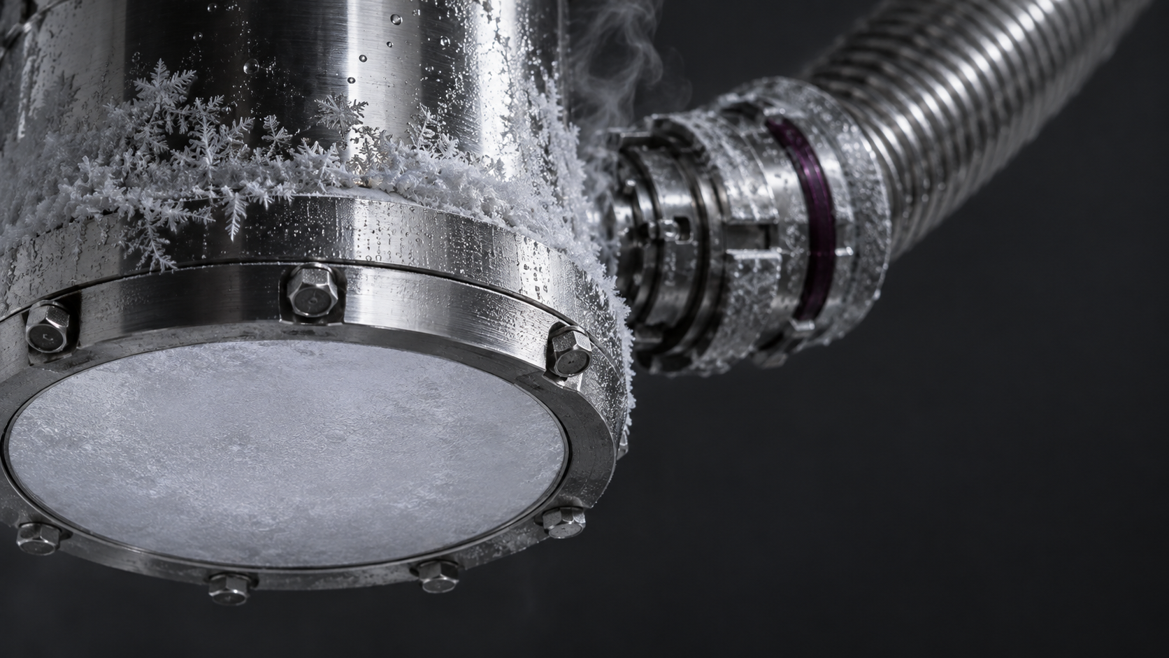
On 4 August 2026 a state press tour called Vibrant China Field Study visited the Institute of Health and Medicine at the Hefei Comprehensive National Science Center and watched a magnetocardiograph acquire signal in the laboratory of Hefei Mandi Medical Technology. Two reports came out of that visit on consecutive days, both from official Chinese outlets, both describing the same machine, and they describe its stage of deployment in different words that no public source resolves.
The company builds a magnetocardiograph around a superconducting quantum interference device, the detector class that reads magnetic fields at the femtotesla scale, and a 2022 industry profile says it obtained an NMPA registration in 2019. The 4 August report describes the machine as working with ten key hospitals in scientific research collaboration and entering a pre-mass-production stage. The 5 August report describes it as installed and in use at more than ten key hospitals in deployed application, with the company's production lines running at full load. A hospital deciding whether to buy one needs to know which of those two sentences is the current state of the product.
A 280-Nanometre Diamond Reached a 682 Microkelvin Resolution Floor: Cambridge, Warwick and Cardiff, 4 September 2026
Quentir Medicine Monitor
Evidence-based insights for quantum medicine.
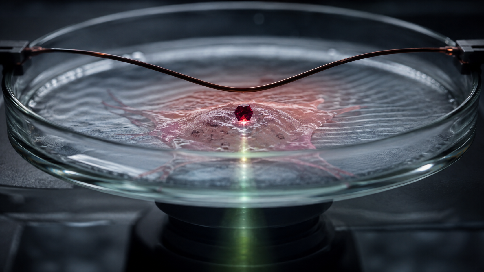
On 4 September 2026 a group drawn from the Cavendish Laboratory at the University of Cambridge, the University of Warwick and Cardiff University posted a preprint on nanoscale thermometry with a free-standing diamond particle. Jack W. Hart, Soham Pal, Julien R. E. Roth, Katie Ninham, Abbie H. Aleksandrova, Xander Peetroons, Soumen Mandal, Oliver A. Williams, Gavin W. Morley, Mete Atature and Helena S. Knowles report that the platform as a whole reaches roughly an order of magnitude better sensitivity than previously published nanodiamond figures.
Three numbers carry the result, and each needs its qualifier. The paper reports an extrapolated resolution floor of 682 microkelvin, a figure derived from the fitting error of the functional form rather than from a single demonstrated reading; the average error on a temperature measurement that took 83 seconds to acquire was 11.9 millikelvin. The sensor is a 280-nanometre nanodiamond ball-milled from isotopically purified carbon-12, small enough in principle to sit inside a cell and holding two nitrogen-vacancy centers rather than one. And the same paper measures the heat its own readout laser pours into the sample, finding 337 millikelvin of heating for every milliwatt of incident optical power, with an observed maximum rise of 224 millikelvin at 0.63 milliwatts, which is some 330 times the extrapolated resolution floor.

IIT Roorkee, 2 September 2026: A Fluorescent-Protein Spin Qubit Six to Eight Orders Short of Neural Nitric Oxide
Quentir Medicine Monitor
Evidence-based insights for quantum medicine.
An individual nitric oxide molecule diffuses about two nanometers in roughly 0.2 nanoseconds, which makes the signal close to its point of production exceptionally transient. The methods available today each miss that measurement in a different way: a microelectrode cannot be placed with nanometer precision, a small-molecule indicator disturbs the redox chemistry it is there to report, and neither reads the radical's own magnetism. On 2 September 2026 five researchers at three Indian institutes published the calculation that says how far one new class of protein sensor sits from doing that job.
The preprint, posted to arXiv as 2609.02792 by Parul Raghuvanshi, Sagnik Ganguly, Sharika E, Mohana Priya T. and Vishvendra S. Poonia of IIT Roorkee, IIEST Shibpur and IIT (ISM) Dhanbad, works out the detection limit for spin relaxometry with the fluorescent-protein spin qubit. Their verdict on the sensor as it exists today is that it misses physiological concentrations of nitric oxide by six to eight orders of magnitude. The bottleneck they identify is the qubit's own relaxation time, which is set by the vibrations of the protein that houses it.
What Feder and colleagues published in Nature on 20 August 2025: a spin-1 qubit inside enhanced yellow fluorescent protein
The object this calculation is about was demonstrated last year. Jacob Feder, Benjamin Soloway and their co-authors reported in Nature 645, 73 (2025) that enhanced yellow fluorescent protein hosts an optically addressable spin-1 qubit in the metastable triplet state of its chromophore. The qubit is initialized by 488 nanometer light through spin-selective intersystem crossing, driven by microwaves, and read out through optically activated delayed fluorescence, in which a 912 nanometer pulse lifts the triplet through a higher triplet state back into the singlet manifold. The delayed light that comes back is separated in time from prompt autofluorescence, so the emission carries the spin state.
Its Hamiltonian is a spin-1 with zero-field splitting, measured at D of 2.356 gigahertz and E of 0.458 gigahertz, which puts three zero-field magnetic resonance lines at roughly 0.92, 1.90 and 2.81 gigahertz. The property that matters for medicine is not spectroscopic. Because the sensor is a protein, a cell can be instructed to build it, and it can be fused to a chosen partner protein. That places the sensor spin within the three-nanometer barrel of an enzyme that makes the very radical one wants to measure. This Monitor read the wider case for cells that grow their own quantum sensors on 26 August 2026; the paper now under discussion is the first hard test of whether the idea survives contact with numbers.
Nanodiamond Charge Drift Reads Macrophage Inflammation: Chicago and Iowa in Advanced Materials, 4 February 2026
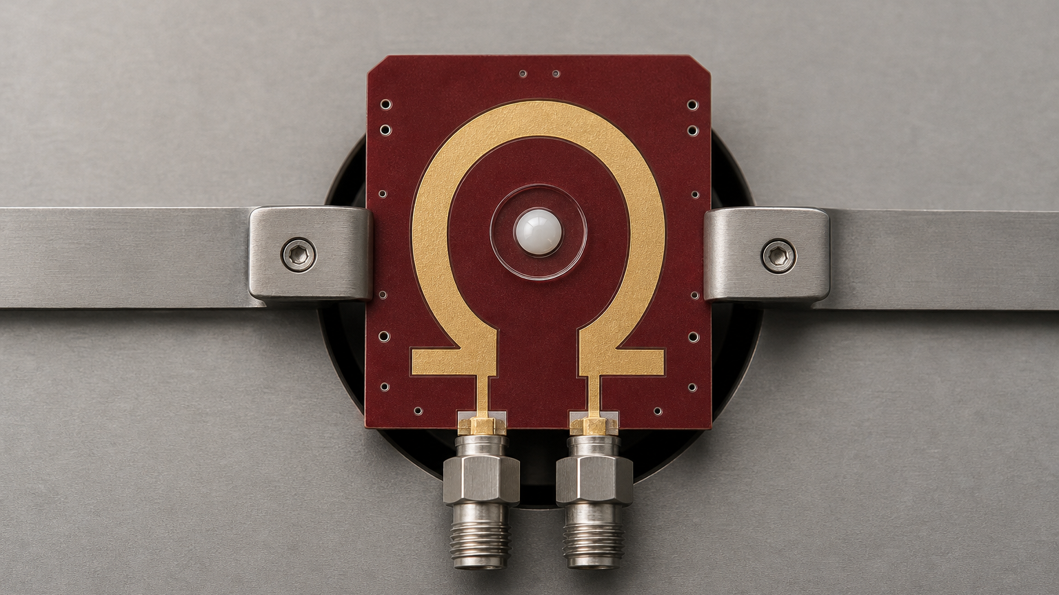
Nanodiamond quantum sensors have been read as thermometers inside living cells for more than a decade. One of them, a 70-nanometer crystal sitting inside a mouse macrophage, drifted downward by 0.27 megahertz over 200 seconds of measurement, which on the usual arithmetic means the cell warmed by 3.62 degrees Celsius. A team at the University of Chicago and the University of Iowa has now put a different quantity behind that same number.
Their paper, Probing cellular activity via charge-sensitive quantum nanoprobes, appeared in Advanced Materials on 4 February 2026, with Uri Zvi as first author and Denis R. Candido at Iowa, Aaron Esser-Kahn and Peter C. Maurer at Chicago as corresponding authors. It was received on 15 March 2025 and accepted on 15 January 2026. The University of Iowa publicized it on 26 August 2026, six months after the article went online, which is why it reaches this Monitor now.
What the zero-field splitting actually tracks, and the dipole term that was being dropped
The sensor is a nitrogen-vacancy center, an atomic defect in diamond whose spin can be initialized with green light, driven with microwaves and read out optically. Each 70-nanometer crystal used here carries roughly 100 of them. The measured quantity is the zero-field splitting, the frequency gap between the spin's ground sublevels, which sits near 2.87 gigahertz and moves by about 74 kilohertz for every kelvin. That temperature coefficient is what turned these particles into intracellular thermometers in the first place.
Cells That Grow Their Own Quantum Sensors
A quantum sensor the cell builds for itself
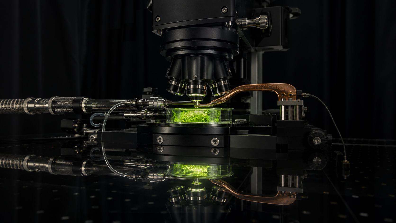
On August 25, 2026 the United States National Science Foundation renewed the University of Chicago-led Quantum Leap Challenge Institute for Quantum Sensing for Biophysics and Bioengineering with a 37.5 million dollar, five-year cooperative agreement running from September 1, 2026 to August 31, 2031, with Chicago State University, the University of Illinois Chicago and Harvard University as partners. The scientific bet underneath the renewal is that a genetically encoded quantum sensor can be written into a cell's DNA and manufactured by the cell itself, rather than fabricated externally and delivered into the cell from outside.
Why genetic encoding matters here
Nanoscale quantum sensing in living systems has until now meant the nitrogen-vacancy center, an atomic defect in diamond that is an exquisite magnetometer and thermometer but arrives wrapped in a lump of diamond that must be introduced into the cell. That is the delivery problem: the particle settles where the cell's machinery leaves it, and steering one to a chosen protein on a chosen membrane is difficult work in its own right. Fluorescent proteins solved the equivalent labeling problem for microscopy thirty years ago without hardware, because they are genetically encodable. Work at Chicago showed that a fluorescent protein can also be operated as an optically addressable spin qubit, roughly ten times smaller than a diamond sensor.
What the record actually shows
The underlying result, published in Nature on August 20, 2025, realized a spin qubit in enhanced yellow fluorescent protein with up to twenty percent spin contrast on near-infrared triggered readout and a sixteen microsecond coherence time under standard decoupling, characterized near eighty kelvin. Three demonstration temperatures have to be kept apart: the full qubit characterization at liquid-nitrogen temperature, coherent control inside human embryonic kidney cells at one hundred and seventy-five kelvin, and optically detected magnetic resonance in living bacteria at room temperature with contrast up to eight percent. No coherent control was shown at human physiological temperature. The authors name photobleaching as the principal limitation and state that sensitivity still falls short of bulk-diamond nitrogen-vacancy sensors, and targeted fusion-protein sensing was not investigated in this study. This Monitor places the work at TRL 3 of 9: real physics, demonstrated in cells, with no clinical measurement and no animal or human study behind it yet.
The Fingerprint Band, Read by a Camera That Never Sees It
A chemical map without a dye
Infrared light between roughly 6 and 10 micrometres is absorbed by the specific chemical bonds that hold proteins, lipids and nucleic acids together, which makes the mid-infrared fingerprint band the part of the spectrum where biological material is most distinguishable without stains, antibodies or fluorescent labels. The reference protocol for the method describes it as a non-perturbative, label-free way to extract biochemical information aimed at diagnosis and at assessing how cells are functioning, and the sample survives the measurement. It has never become routine hospital equipment, and one important barrier is the detector rather than the chemistry.
The camera problem, answered sideways
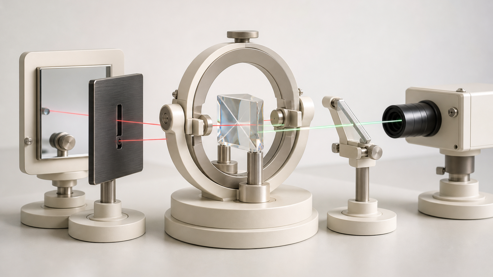
Detectors for that band are cooled, costly, and limited by the thermal glow of the room and of the instrument itself, because everything at ordinary temperature radiates in exactly the wavelengths being measured. A group at Imperial College London has now reported wide-field imaging across the full 6 to 10 micrometre range in which the infrared light is never measured at all. Correlated photon pairs are produced in a single silver thiogallate crystal used twice in a folded geometry, one partner passes through the sample, and the picture is reconstructed from the visible partners of those undetected photons on a commercial scientific silicon camera. Because the measurement happens in the visible, where the room's thermal background is effectively absent, the system detects infrared signals about a hundred times below the usual background-limited photodetection ceiling, at room temperature.
What the numbers actually allow
At 8 micrometres the images hold more than 8,000 resolvable elements at a resolution of 297 plus or minus 5 micrometres, over a circular field about 30 millimetres across, in a 10 second acquisition. The field is generous enough for a tissue section, a tablet or a culture well. The resolution is the constraint that matters: a human cell measures 10 to 20 micrometres, so that resolution is equivalent to roughly fifteen to thirty cell widths rather than to a camera pixel of that size, and the test objects were shadow masks cut from metal foil rather than biological material. This edition reads the result as a supply-chain and noise-floor contribution to an established clinical method, places it on the readiness ladder, and sets out the three specific demonstrations that would tell a hospital buyer the distance to a pathology bench is closing.
A Diamond Magnetometer Closes the Distance to Biomagnetism
A smaller gap between sensor and signal
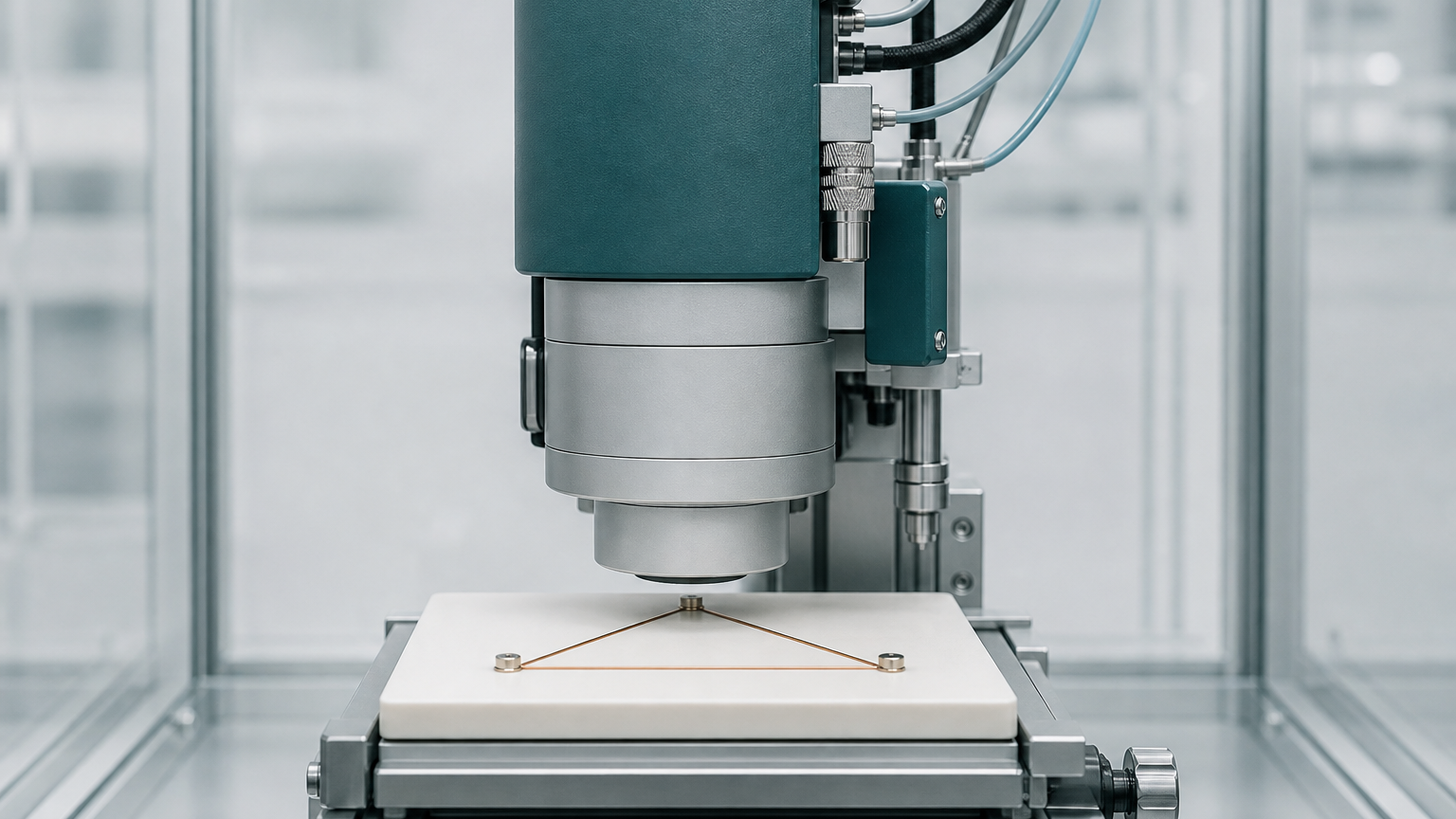
Biomagnetic fields weaken sharply with distance, so the physical gap between a detector and the body is part of the sensitivity budget. Yuta Araki and colleagues have built a diamond NV magnetometer whose sensing head can sit about 2.0 millimeters from a sample. A compact microwave antenna helps set that geometry, while a light-trapping diamond waveguide makes more efficient use of the green laser that initializes and reads the sensor's nitrogen-vacancy centers. The integrated device addresses the heat and bulk that have limited high-sensitivity Ramsey measurements near biological samples.
What the laboratory result establishes
The peer-reviewed paper reports 2.93 picotesla per square-root hertz sensitivity across 100 to 400 hertz at 210 milliwatts of laser power. The measured temperature increase was approximately 13 kelvin. In a controlled test, the sensor detected a 77.7-picotesla field from a dry brain-field phantom at a 2.5-millimeter standoff, without signal averaging and with a signal-to-noise ratio of approximately 4.3. Quentir assesses the assembled system at TRL 4 of 9: a quantum sensing device validated in the laboratory on a phantom that imitates a brain-field pattern.
The clinical distance remains
The phantom does not reproduce movement, anatomy, variable spacing or the environmental interference of a living-subject recording. The authors also state that sub-picotesla brain signals will require further accumulation and improved sensitivity. A future magnetoencephalography or magnetocardiography instrument would need stable arrays, calibration and clinical comparisons as well. The current paper supplies no human dataset, workflow study, regulatory record or manufacturing claim. Those absences keep the result at the laboratory-instrument stage even though its geometry addresses a genuine near-body constraint. Quentir reads the paper as a bounded hardware advance. It joins optical efficiency to a short sensor-to-sample distance, reports each operating constraint quantitatively, and makes the next test easy to name: a living-subject measurement with a defined physiological signal and an established comparator.
What Yonsei's Metabolic MRI Installation Has to Prove
A university project gets a deployment window
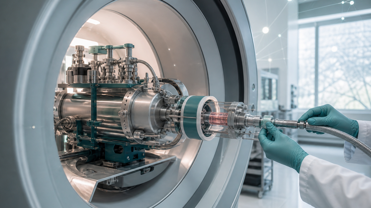
The RESQ consortium plans to install NVision's POLARIS platform at Yonsei University in early 2027. Backed by €2,572,500 in Horizon Europe funding, the three-year program joins NVision, Yonsei University, Ulm University, and Tesla Dynamic Coils. Together they are building a metabolic MRI installation for higher-resolution research on brain metastases. The public project record assigns concrete work: reproducible parahydrogen generation and storage, tailored radiofrequency pulses, compressed sensing, machine learning, and dedicated dual-tuned brain coils. These details turn a broad quantum-health ambition into a dated engineering program with named owners.
The scanner is only one part of the workflow
Hyperpolarization can strengthen the MRI signal from selected metabolic agents for a limited period, allowing researchers to study what tissue is doing alongside its anatomy. That advantage can disappear if gas preparation, sample transfer, coil performance, pulse design, or image reconstruction varies. RESQ therefore has to integrate chemistry, hardware, software, scanner time, and operating procedure. Its stated goal of doubling spatial resolution is a consortium target. The current public record does not report a completed Yonsei installation, a disease-model result, a patient study, diagnostic accuracy, or a change in treatment.
Preclinical work is the next meaningful test
Yonsei is expected to validate the workflow using preclinical brain metastasis models. That stage can show whether the complete system produces stable metabolic maps in biologically relevant models and whether smaller lesions become more visible. It cannot yet establish performance in patients. Quentir reads RESQ as a quantum-sensing implementation program at TRL 4 of 9: the underlying hyperpolarization platform operates in research settings, while the new disease-specific workflow still awaits installation and integrated preclinical validation. The decisive question for the project period ending in April 2029 is whether preparation, acquisition, coil behavior, and reconstructed images can be reproduced beyond one expert site. A portable workflow would move quantum sensing closer to useful medical infrastructure; a result dependent on one installation would leave clinical translation much farther away.
The MRI Implication of a Single Copper-Oxide Plane
The result is one atomic plane
A team led by researchers at Fudan University and the University of Science and Technology of China has fabricated a single-layer cuprate containing one superconducting copper-oxide plane. Published in Nature on August 12, the experiment reduces Bi-2201 to its ultimate two-dimensional limit. The monolayer retained superconductivity, although its optimal transition temperature was about ten percent lower than in thicker material. Fine control of oxygen content then let the researchers follow the sample from an insulating state, through an anomalous metallic regime, and into superconductivity. This is a basic-physics achievement built on delicate fabrication and a laboratory instrument designed for precise in-situ tuning.
The medical relevance begins with the magnet
MRI depends on a strong, stable magnetic field to align protons in the body before radiofrequency pulses and sensors turn their response into anatomical images. That creates an MRI materials pathway for this research, but the connection sits far upstream. The paper reports no magnet winding, imaging coil, scanner prototype, patient study, or medical-device test. Its contribution is a cleaner experimental platform for understanding how high-temperature superconductivity changes when the active material is reduced to one copper-oxide plane.
The distance to care remains useful to measure
A hospital magnet requires far more than a superconducting transition. Engineers need scalable conductors, high current under strong fields, reliable joints, mechanical strength, controlled cooling, quench protection, field homogeneity, and compatibility with a complete scanner. The new monolayer does not answer those engineering questions. It helps make the earlier materials questions more exact. Quentir reads the result as a sensing story at the laboratory-materials stage: credible MRI relevance through superconducting magnet science, paired with a clear boundary around present readiness. The next meaningful step would connect the one-plane physics to a thicker, manufacturable material with measured current or field performance.
A One-Number Test for the Brain's Signal Carriers
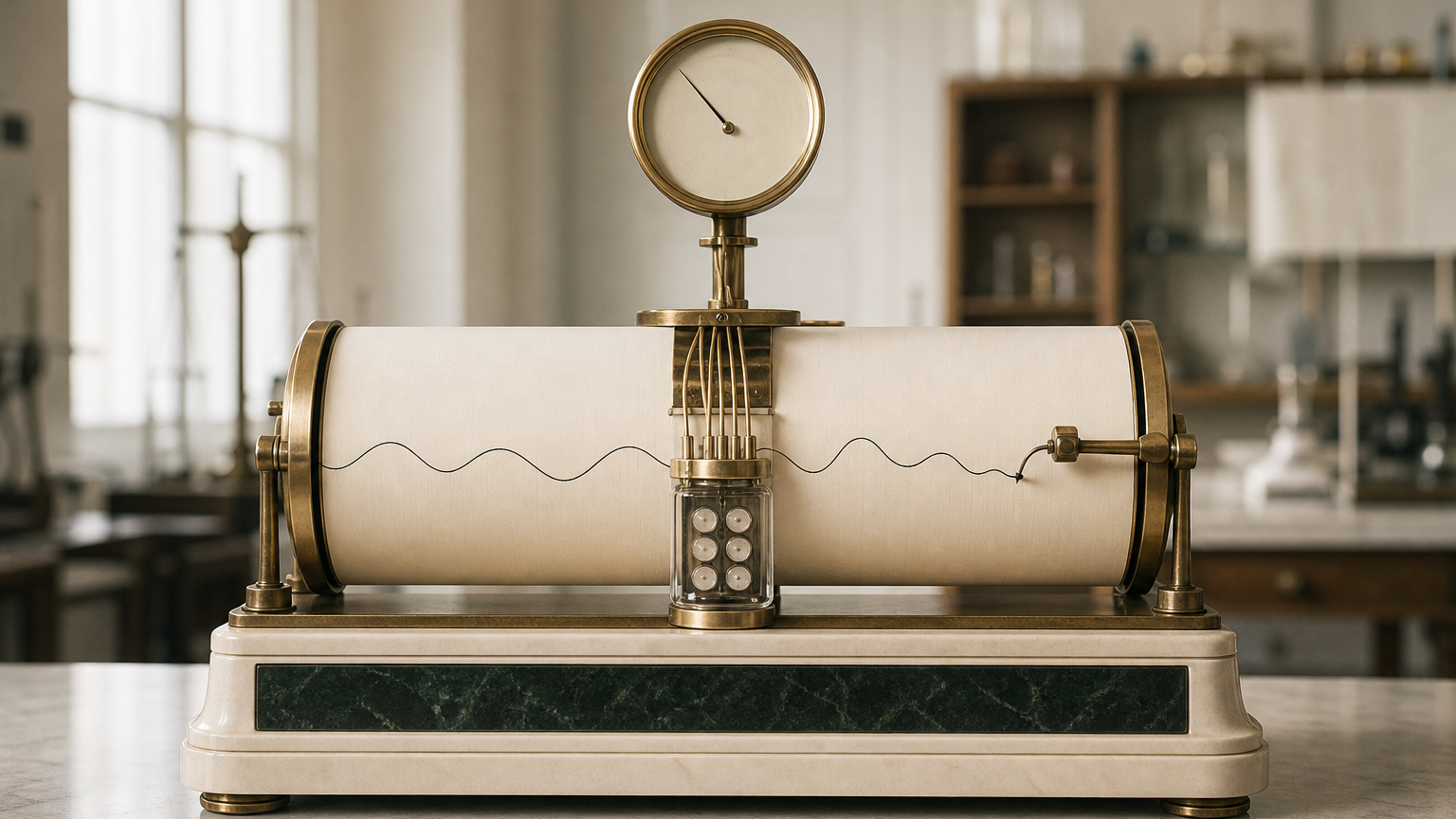
One number bounds what a brain signal can carry
A preprint posted to arXiv on August 11 gives the quantum-brain debate something it has lacked: a shared standard. Eran Kopel shows that spectral distinguishability alone bounds the labels a biological oscillator can carry by its quality factor, two pi times frequency times coherence time. The ceiling holds for any substrate and any mechanism, takes no position on quantum effects in biology, and can be computed from two published quantities. Collective vibrational modes, endogenous electromagnetic fields, microtubule excitations and oscillatory phase codes now face one arithmetic instead of four separate arguments.
A proposed cortical microwave field fails by every route
Applied to a recently proposed 30 gigahertz field inside cortical columns, the screen returns a quality factor of 0.19, a linewidth five times the carrier frequency. The rescue of a driven, spectrally narrow emitter requires a resonant cavity the model's own geometry forbids, and an independent metabolic-power bound is exceeded by five to nine orders of magnitude. Of eleven screened carriers, only the low-frequency neural rhythms pass. High-frequency molecular carriers fall to brevity, and the fragility argument the debate assumed proves unnecessary.
Braintech rides the rhythms that pass
Closed-loop EEG platforms already work in the frequency range the screen favors, portable designs keep maturing, and national programs are putting dates on commercial neurotechnology. Quentir reads the screen as inexpensive governance for an expensive decade: state the frequency, state the coherence time, and accept the ceiling they imply, with quantum sensing as the measurement arm being developed to pin those numbers down in living tissue.
Australia Funds a Quantum Window Into Living Brain Tissue
The project begins before the clinic
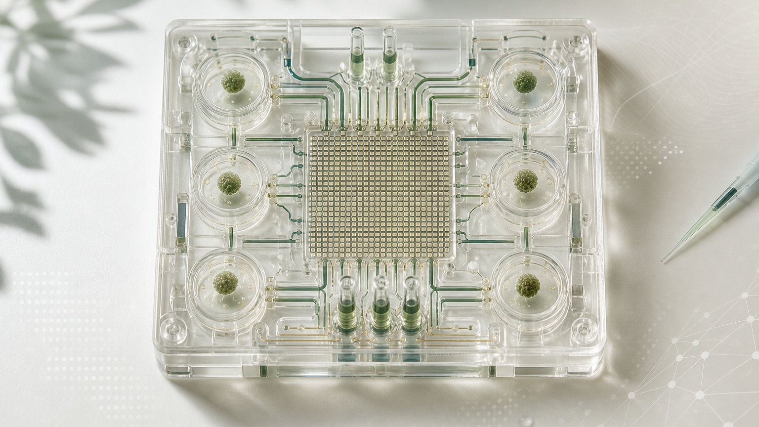
Australia has awarded AUD 2.1 million to a University of Melbourne consortium developing a brain-on-chip platform for neurological drug research. The partners are Chromos Labs, Tessara Therapeutics, Quantum Brilliance, and Axol Biosciences. Their stated plan joins human neural micro-tissue to quantum tools that measure electrical activity in real time. The target is preclinical work: observing how laboratory-grown neural tissue responds to candidate therapies before a claim reaches patients. The University announcement names Alzheimer’s disease, schizophrenia, epilepsy, and anxiety disorders as intended fields of use. A later Team France Export account repeats that list and explicitly credits the University source. The disease names describe ambition, not validation. The public record reports a funded development program and no integrated-system result or clinical study.
Electrical behavior can reveal what a final endpoint misses
Neurons communicate through changing electrical states. Continuous measurement can show when a tissue model responds, how long the response lasts, and whether activity returns to baseline. That makes the platform relevant to preclinical drug discovery, where an earlier rejection of a weak candidate can save years of work and reduce the chance that patients enter a trial built on a fragile signal. The benefit depends on control. Tissue batches vary, microfluidic conditions drift, and sensitive electronics can mistake ordinary noise for a drug effect. A useful platform will need blinded comparisons, repeatable preparation, reference measurements, and a clear path from raw signals to the claimed biological response.
The quantum component still needs a technical record
The project accounts describe quantum technology as part of the real-time measurement layer. They do not disclose the sensor architecture, detection limits, noise floor, calibration method, or comparison instrument. That keeps the readiness answer open. Quentir classifies the function as quantum sensing while withholding a numbered readiness level from a grant announcement with no reported integrated result. The next credible milestone is an assembled laboratory system tested on human neural micro-tissue, with controls that distinguish a drug response from sensor noise, tissue variability, and software choices. If that milestone lands, the project may become a better filter for neurological drug pipelines long before it becomes anything a patient or hospital encounters directly.