A 90-Degree Antenna for a Difficult Organ
Quentir Medicine Monitor
Evidence-based insights for quantum medicine. Published by Quentir Systems LLC · August 4, 2026.
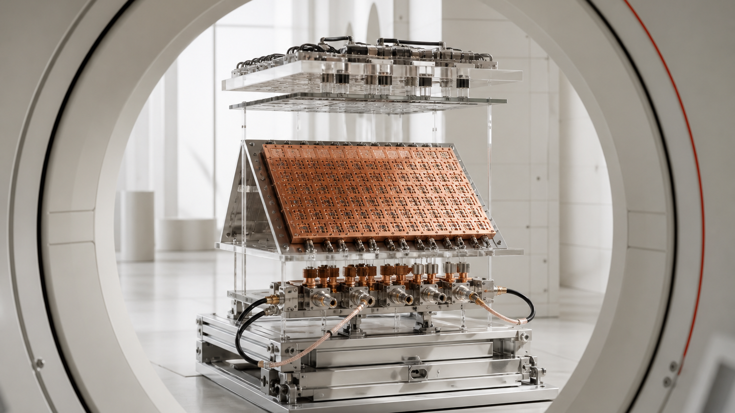
The human eye is a difficult target for magnetic resonance imaging. It is small and never perfectly still during a scan; nearby tissues also respond differently to a strong radiofrequency field. Higher magnetic fields can reveal finer anatomy, yet the physics that creates more signal can also produce shading and signal voids. Local heating adds a separate safety concern.
A team in Berlin and Rostock built a metamaterial antenna around that problem. Its printed circuit board bends through 90 degrees over the eyes, like a shallow gable roof. Forty subwavelength copper cells sit inside the antenna structure and reshape the near field at the 297.2-megahertz operating frequency of a 7-tesla scanner.
The result is a compact piece of hardware for ocular MRI, tested in phantoms, five healthy adults, and one person with treated retinal disease. The antenna increased transmit efficiency and received signal compared with a matched loop design. It also illustrates a boundary that quantum medicine needs to keep clear: the scanner reads quantum spin behavior, while the metamaterial itself is an engineered radiofrequency structure.
Practical takeaway. This is a human feasibility study of a new 7-tesla MRI antenna. It supports further device development for eye and posterior-brain imaging, but it does not establish diagnostic superiority, regulatory readiness, or routine clinical use.
Why the eye is a hard MRI target
Ophthalmic MRI has an unusual combination of demands. The anatomy is delicate and measured in millimeters. The eye moves even when a person is trying to keep still, so long acquisitions invite blur. Ultrasound and optical imaging already serve many eye-care questions, while MRI is useful when clinicians need soft-tissue contrast, deeper orbital coverage, or a view behind opaque structures.
Moving from 3 tesla to 7 tesla can improve spatial resolution and sensitivity. It also shortens the radiofrequency wavelength in tissue. That can make the transmitted field uneven, leaving dark regions or bright patches in an image. Radiofrequency energy deposition and tissue heating also matter more at high field, especially around a conductive and temperature-sensitive organ.
The peer-reviewed study by Nandita Saha and colleagues treats the antenna as part of the imaging material. Each of its forty cells is a double-square split-ring resonator. The cells are smaller than the operating wavelength and arranged in a five-by-eight array on the same substrate as a two-channel transmit-and-receive loop. Near-field coupling between the layers changes how energy reaches the tissue and how the returning signal reaches the receiver.
The geometry is clinically legible. A flat version sits behind the head for the occipital lobe. A second version bends at a right angle and rests above the anterior head without touching the face. The researchers compared each metamaterial design with a conventional loop built to the same dimensions and tuned to the same frequency.
Quantum pillar: sensing. Technology readiness: TRL 6 of 9. A working prototype completed realistic eye and brain scans in healthy volunteers and one person with retinal pathology, but it has no regulatory clearance or routine-care record.
What the antenna changed in living subjects
The direct human comparison involved three healthy volunteers scanned with both the bent metamaterial antenna and the matched bent loop. Across the two eyes, transmit-field gains ranged from 10 percent to 40.2 percent. T2-weighted imaging showed signal gains of 25 to 51 percent in the left eye and 26 to 29 percent in the right. T1-weighted gains were smaller, from 7 to 26 percent, but remained positive across the reported comparisons.
Those percentages are encouraging and narrow. They describe regions of interest in a small feasibility cohort, not sensitivity or specificity for a disease. The study does not compare diagnostic decisions, patient outcomes, examination time, or performance against every coil system a hospital might already own.
The clinical images still matter. Five healthy adults underwent ocular scans with the new antenna. One volunteer returned for four sessions on separate days, giving the team a limited repeatability check. The researchers also imaged a 26-year-old participant with Von Hippel–Lindau disease and a treated retinal hemangioma. The scan showed post-treatment structural change without apparent progression and a separate vascular malformation known from prior assessment. An incidental maxillary sinus cyst was visible as well.
That single case demonstrates anatomical reach, including treated and incidental findings. It cannot establish a diagnostic claim. The patient was scanned 16 days after treatment, and the paper reports no blinded comparison with another modality or reader study. The proper reading is feasibility: the device produced interpretable images in a person whose anatomy gave the prototype a meaningful test.
The flat antenna added a second target. In two healthy volunteers, it improved transmit efficiency by 30 and 44 percent over the planar loop and extended signal coverage across the occipital region. That result connects the same materials architecture to neuro-ophthalmic imaging, although the evidence remains even smaller than the ocular series.
Safety is part of the performance claim
An antenna that intensifies a radiofrequency field near the eye must answer a thermal question. The authors combined electromagnetic simulations, human voxel models, phantom tests, magnetic-resonance thermometry, and fiber-optic temperature probes. In their simulations, local specific absorption rates remained below the cited IEC limits. The highest modeled local value for the bent configurations was about 1.36 watts per kilogram, and the value near the eye remained below 1 watt per kilogram.
A 30-minute, 10-watt heating simulation predicted a maximum local temperature rise of about 0.7 degrees Celsius. Phantom experiments with thermometry and fiber-optic probes supported the modeled spatial pattern. These are useful engineering controls, and the participants received standard 7-tesla safety procedures and post-scan interviews.
Hospital buyers will still need a later evidence package. Safety depends on the final device and scanner platform, together with its sequences and operating limits. Manufacturing tolerances and patient anatomy matter too. Intended use sets another boundary. A prototype that stays within limits in a controlled study has crossed an important threshold. It has not completed qualification.
The commercial path also deserves ordinary disclosure. The paper states that coauthor Thoralf Niendorf is founder and chief executive of MRI.TOOLS GmbH, which appears in the hardware chain described by the study. The project received European Research Council and German Research Foundation support. The underlying participant data are available from the corresponding author on request and are not public because of privacy and ethics restrictions.
Quantum materials and quantum medicine are not synonyms
The paper calls its device an electromagnetic metamaterial. Its double-square cells produce an engineered negative-permittivity response and shape the radiofrequency near field. No quantum computer or quantum algorithm appears in the experiment.
MRI nevertheless belongs to the sensing pillar because its signal comes from nuclear spin and magnetic resonance. The antenna controls the classical radiofrequency field used to excite and receive that quantum-derived signal. Both layers matter, and they should not be collapsed into one label.
A July 30 account from UC San Diego helps explain the wider materials connection. The university's NSF-supported materials center is studying metamaterials whose active building blocks are nanoscale quantum materials. Its article also points to clinical MRI as an existing area in which metamaterials can shape radiofrequency fields and may improve image quality or acquisition time. That context joins two research lines without proving they use the same mechanism.
The distinction is more than terminology. Classical metamaterials can improve a quantum-sensing instrument now. Quantum metamaterials may later offer different active properties and finer control at smaller sizes. A hospital evaluating an MRI antenna needs demonstrated field maps and thermal limits. Human images and the regulatory status of the actual device belong in the same assessment. A broad “quantum material” label supplies none of those facts by itself.
How Quentir Reads It
Quentir reads this paper as a materials-to-device demonstration with unusually complete early engineering. The team designed the unit cell, integrated it with the antenna, benchmarked matched hardware, tested phantoms and human subjects, and treated heating as part of performance. The study also includes one pathological case, which is useful precisely because it remains a case rather than a clinical cohort.
The original connection lies in where the innovation sits. The scanner magnet is the conspicuous capital asset, but the decisive change here is a shaped copper structure close to the body. Medical imaging can advance through the interface that couples a patient to a mature machine. That shifts attention toward anatomy-specific accessories and manufacturing consistency. Cleaning and setup time affect operations; comfort and coil interchangeability affect adoption.
The clinical limit is equally clear. The cohort is small, the comparison is technical, and the device is tied to an ultrahigh-field research environment. Larger studies would need to show reproducibility across operators and body types, compare diagnostic tasks, and determine whether the signal gain shortens scans or changes care. Qualification and serviceability would follow.
For a patient with a retinal mass or an orbital disorder, finer soft-tissue imaging could sharpen treatment planning or follow-up. That humane possibility is why the hardware deserves attention. Today, the paper shows that a carefully shaped antenna can carry the quantum signal more effectively around a difficult organ. Medicine still has to prove what that extra signal changes.
Sources
Primary source: Saha and colleagues, Advanced Materials, published online February 2, 2026. Materials context: Kane and Franklin, UC San Diego Today, July 30, 2026.