IIT Bombay Review, 8 October 2026: Where Diamond Quantum Sensors and Fluorescent-Protein Qubits Stand for Medicine, From Living Cells to a Rat's Heartbeat
Quentir Medicine Monitor
Evidence-based insights for quantum medicine. Published by Quentir Systems LLC · October 9, 2026.
A cardiologist reads the heart through electrodes on the skin, and a neurologist reads the brain through sensors on the scalp. A small research field is trying to place quantum sensors next to the molecules themselves, inside cells and tissue, where they can report temperature, magnetic fields or chemical stress from within.
On 8 October 2026 three engineers at the Indian Institute of Technology Bombay posted a review of where that effort stands. Jyotiprakash Parhi, Ayan Majumder and Kasturi Saha follow quantum sensors for biomedicine from their established form, tiny defects in diamond, to a newer one: the fluorescent-protein qubit, a sensor a living cell can make for itself from its own genetic instructions. Between those two ends sit the nanodiamond probes that have already been placed inside cells to measure heat and the stiffness of the material around them.
The paper, "From Solid-State Spin Defects to Fluorescent Proteins: A Perspective on Quantum Optical Sensing Platforms for Biomedical Applications," is a preprint on arXiv (2610.11644) and has not been peer reviewed. It reports no new experiment. Its value for a hospital research director, a pathologist or a medical-device buyer is the map it draws: which measurements have been shown in cells, which in a living animal, and which problems the authors say still stand between these sensors and clinical use. A summary table sets out, sensor by sensor, the furthest biomedical stage reached, the representative study and the main barrier to translation.
The common thread is a defect or molecule whose electron spin changes how brightly it glows. Shine green or blue light on it, often together with a weak microwave signal, and the glow dims or brightens depending on the magnetic field, the temperature or the chemistry nearby. Because the readout is light, a microscope can collect it from inside a cell without wires.
What Parhi, Majumder and Saha at IIT Bombay compared in arXiv 2610.11644
The review covers four families of sensor. The most widely studied is the nitrogen-vacancy center, a missing carbon atom next to a nitrogen atom in the diamond lattice, which works at room temperature. The silicon-vacancy center, another diamond defect, gives a sharp color line that shifts with temperature and allows thermometry by light alone. Boron-vacancy defects in hexagonal boron nitride, a flat material a few atoms thick, extend the idea to surfaces that can be laid directly under cells. The fourth family is biological: proteins whose light-activated states carry an electron spin that can be read out optically.
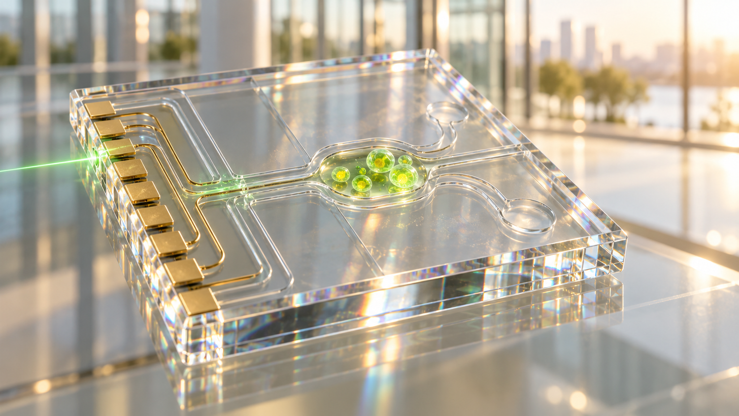
The authors then sort published experiments by scale: single molecules, the inside of cells, and tissue. They also cover the engineering that carries a sensor toward a usable device, including diamond photonic structures that collect more light, chips built for cell culture and animal work, microfluidic channels that keep samples in controlled liquid, and diamond nanoneedles that touch a cell with minimal damage.
Which diamond sensors already work in cells and in a living rat: 100 millikelvin thermometry and magnetocardiography
Inside living cells, the review lists nanodiamond measurements of temperature with a precision down to about 100 millikelvin, maps of the viscosity of the cell interior, and relaxometry, a method that senses free radicals through the magnetic noise they produce. One study it cites, by Mzyk and colleagues in Advanced Functional Materials in 2026, used nanodiamonds to follow free-radical and iron changes in human cardiac fibroblasts as the cells moved toward a scar-forming state, a cell model of cardiac fibrosis. The review adds the caution the evidence calls for: further work in tissue and in animals is needed before such signals could serve as biomarkers of fibrosis. Beyond single cells, its summary table lists nanodiamond detection of free radicals in slices of mouse liver and nanodiamond temperature measurement inside the tissue of a living rat.
A separate animal demonstration comes from a 2024 study in Physical Review Applied by Yu and colleagues. They recorded the magnetic field of a beating heart in a living rat with a diamond sensor at room temperature, without contact, reaching a sensitivity of about 9 picotesla per square-root hertz with the help of magnetic flux concentrators. Clinical magnetocardiography has mostly relied on SQUID sensors cooled with liquid helium and, more recently, on optically pumped atomic magnetometers; a sensor that works at room temperature is one route toward smaller and cheaper equipment.
Quantum pillar: sensing. Technology readiness: TRL 3 of 9, rated for one application: nanodiamond temperature sensing inside living tissue, which has reached a proof-of-concept measurement in a rat and no study in patients. External diamond heart recording has likewise reached a single rat study, and the protein sensors have so far worked only in cells and bacteria.
How a University of Chicago team built a protein qubit and an Oxford-led team engineered magnetic-sensing proteins, in Nature in 2025 and 2026
Diamonds must be delivered into a cell from outside, and their surfaces have to be coated so the cell tolerates them. The review's central argument is that a sensor made by the cell itself removes that step. Two papers carry it. In a Nature paper published online on 20 August 2025, Jacob Feder, Benjamin Soloway and colleagues at the University of Chicago and Argonne National Laboratory turned enhanced yellow fluorescent protein, a standard label in biology laboratories, into a spin qubit. At liquid-nitrogen temperatures they read it out with up to 20 percent contrast and kept it coherent for about 16 microseconds. They expressed it in mammalian cells, and in bacterial cells they measured its magnetic resonance at room temperature with contrast up to 8 percent.
In Nature in January 2026, Gabriel Abrahams and colleagues, led from the University of Oxford, reported engineered magnetic-field-sensitive proteins called MagLOV. These work through a different mechanism from the yellow protein qubit: a short-lived radical pair formed between the protein and a bound flavin cofactor. Through directed evolution they changed how the proteins respond to magnetic fields and radio waves, detected magnetic resonance in living bacteria at room temperature with enough signal for single cells, and used field gradients to locate the glow in space, a form of magnetic resonance imaging with a genetically encoded probe. The Monitor examined the distance between a fluorescent-protein qubit and a neural signal in September; the IIT Bombay review places both Nature papers in the wider set of tools.
Why surface noise, protein folding and tissue scattering still keep these sensors in the laboratory
The review is direct about limits. For diamond sensors near the surface, stray magnetic fluctuations from dangling bonds, adsorbed molecules and surface atoms shorten the time the spin holds its state and reduce sensitivity. The charge state matters too: only the negatively charged defect senses, and surface chemistry can switch it off. Coatings that let a diamond bind a target molecule can push the target further away and weaken the signal, so the surface chemistry has to serve the biology and the physics at once.
Proteins bring their own problems. They are packed with atomic nuclei whose small magnetic fields limit how long the spin stays coherent, and folding, the surrounding water and changing conditions inside the cell make the signal vary from one setting to another. At the scale of tissue, light scatters in thick samples such as liver slices, the body's own iron and other magnetic material blur specific signals, and particles spread unevenly. The authors conclude that reproducible calibration across conditions is a prerequisite for quantitative use.
What hospital research teams and device buyers can take from the review in October 2026
For a clinical audience the practical reading is about time and setting. Diamond thermometry and relaxometry are research instruments for cell biology and drug studies, used by specialist groups with custom optics. In living animals, the review cites a room-temperature diamond heart recording from outside the body of a rat and nanodiamond thermometry inside rat tissue, both far from a validated diagnostic test. Fluorescent-protein qubits are about a year old as a published platform and have so far worked best in bacteria at room temperature.
The direction the review describes is clear: sensors that a cell grows for itself, chips that hold living samples, and diamond probes that report from inside a single cell. The next evidence worth watching is a fluorescent-protein sensor reading a medically relevant signal in mammalian cells at body temperature, and a diamond heart or nerve recording repeated in a larger animal by an independent group.
Sources
Primary source: Jyotiprakash Parhi, Ayan Majumder and Kasturi Saha, From Solid-State Spin Defects to Fluorescent Proteins: A Perspective on Quantum Optical Sensing Platforms for Biomedical Applications, arXiv preprint 2610.11644, 8 October 2026. Also drawn on: Feder et al., Nature, 2025; Abrahams et al., Nature, 2026; and Yu et al., Physical Review Applied, 2024. Readiness and implications are the Monitor's editorial assessments.