Quentir Medicine Monitor
Evidence-based insights for quantum medicine. Published by Quentir Systems LLC · October 31, 2026.
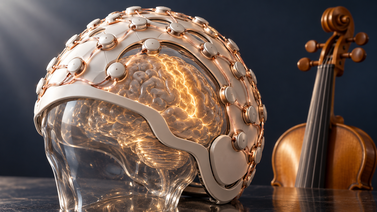
Physicists at the University of Nottingham have recorded the brain activity of 22 adults while each one learned to play a violin, using a wearable helmet of optically pumped magnetometers that moved with the head. The study shows that this form of OPM-MEG can capture usable brain rhythms, after noisy trials are removed, while a seated person plays an instrument with head, neck, shoulder and arm movement, a task the paper calls difficult or even impossible in the fixed, cryogenically cooled scanners of hospital MEG suites.
The paper appeared online in the open-access journal Imaging Neuroscience on 22 October 2026, and Optics.org reported it on 29 October 2026. Its central finding concerns beta oscillations, the electrical rhythms of the brain between about 12 and 30 Hz: after a single 30-minute lesson, beta power in the motor and pre-motor areas rose while the participants played. Beta rhythms are disturbed in Parkinson's disease, multiple sclerosis and other conditions, so a scanner that records them during ordinary movement is of direct interest to neurology departments.
Matthew Brookes, who leads OPM-MEG development at Nottingham and chairs the spin-out company Cerca Magnetics, told Optics.org that some of the effects measured in the study are among those that can be affected in disorders like dementia. The study itself tested healthy volunteers only.
What Joseph Gibson, Matthew Brookes and nine colleagues published in Imaging Neuroscience
The peer-reviewed article, titled "Beta-band dynamics during a naturalistic motor task: An OPM-MEG study", is article IMAG.a.1405 in volume 4 of Imaging Neuroscience, published under a Creative Commons Attribution license. Joseph Gibson is the first author and Matthew J. Brookes the senior author. Ten of the eleven authors are listed at the University of Nottingham's School of Physics and Astronomy, and Matias Ison at the university's School of Psychology. The manuscript was received on 23 December 2025 and accepted on 31 August 2026.
The experiment had a simple structure. Each participant watched a short video of an expert violinist playing the first four bars of "Twinkle Twinkle Little Star", then tried to play the tune while the scanner recorded. The same expert then gave a lesson of about 30 minutes covering how to hold the violin and the bow, open-string bowing, finger spacing and the notes. A second scanning session followed, and each session lasted about 35 minutes.
The team had to build a violin with no metal parts. Moving metal near the head creates magnetic artifacts, so the researchers fitted gut strings, a bow strung with natural horsehair and wooden fittings, removed the metal fine-tuners and retuned the instrument to baroque pitch, with the A string at about 415 Hz.
Quantum pillar: sensing. Technology readiness: TRL 6 of 9. On Quentir's assessment, the wearable system works reliably on human volunteers inside a magnetically shielded research room, and research versions are already sold to neuroscience centers, while routine use for diagnosing patients in hospital care has not yet been established.
How 64 rubidium vapor sensors measure the brain's magnetic field without liquid helium
Conventional MEG scanners use superconducting sensors fixed in a rigid helmet and cooled with liquid helium, so the patient has to keep still. Each optically pumped magnetometer in the Nottingham helmet instead holds a small glass cell of rubidium-87 vapor, a laser and a photodiode. The laser prepares the quantum spin state of the rubidium atoms, and the tiny magnetic fields produced by brain currents change how much light passes through the cell. The paper gives the size of each sensor as 12.4 by 16.6 by 24.4 millimeters and its weight as about 4 grams.
The system used 64 triaxial sensors from the US manufacturer QuSpin, each sensitive to the magnetic field in three directions, for 192 measurement channels across the scalp. The sensors ran in a closed-loop mode that kept their output linear while the head moved. The whole setup sat inside a 3 by 3 meter shielded room lined with four layers of mu-metal and one of copper. The same family of vapor-cell magnetometers appears in other settings: Quentir has covered a 7 cubic centimetre rubidium magnetometer field-tested by USTC researchers for navigation and detection work.
The Nottingham group first showed a wearable OPM system in a Nature paper of March 2018, in which participants could move their heads during a scan. Because the sensors can be placed close to any scalp, the approach also suits small heads, and the group has used it with children, a line of work Quentir followed in its report on how the helmet is bringing brain mapping closer to childhood.
What the beta rhythm measurements showed before and after the 30-minute violin lesson
The recordings behaved as the field expects for movement. Beta amplitude dropped while participants played and rose again when they stopped. After the lesson, beta power during playing was significantly higher in the motor and pre-motor areas than before it, and the difference between playing and rest became more pronounced. These two results fit together because beta power at rest was also significantly higher after the lesson, so a larger dip during playing started from a higher baseline.
The authors read this through predictive coding models of the brain, in which beta amplitude rises when a person has more certainty about the movement being made. Gibson described the idea to Optics.org in plain terms: the brain expects a certain sound from each bow stroke and registers whether the note matched the prediction. The study measured these signals while the instrument was being played, which is the methodological point for clinicians.
What a neurology department or hospital buyer should note about the limits of this study
The authors state the main technical limit themselves. The helmet ran without the large field-cancelling coils that normally suppress the background magnetic field, because the motion-capture system already controlled the experiment and adding field control was difficult. Head movement in the residual field therefore still produced artifacts, and the team removed noisy trials by visual inspection. The authors judged this acceptable for beta rhythms, which sit at relatively high frequencies in healthy adults, and the same choice would be harder to defend for slower rhythms or for patients who move more.
The sample was 22 healthy adult novices, the task was one tune, and the follow-up came minutes after a single lesson. The study therefore shows that the technology can follow a learning brain during natural movement. It does not show that beta measurements can diagnose dementia, Parkinson's disease or any other condition, and no patients took part. The participants rated their own playing on a five-point scale, and the links the team found between beta changes and improvement in those ratings did not survive correction for multiple comparisons, so the authors report them only as a trend. They also note that muscle fatigue may have affected the second session more than the first.
Readers should also weigh the commercial link and the regulatory position. Cerca's website states that its OPM-MEG system is offered as a research system and does not have any medical or regulatory approvals in any jurisdiction. According to the paper's declaration of competing interests, four authors hold founding equity in Cerca Magnetics, and Elena Boto and Matthew Brookes are directors of the company. Optics.org reports that Cerca raised £3.8 million in Series A funding in April 2026 and had sold 19 research systems to neuroscience centers in twelve countries. The work was funded by the Engineering and Physical Sciences Research Council, the UK Quantum Technology Hub in Sensors, Imaging and Timing, and a Medical Research Council equipment grant.
How Quentir reads the Nottingham violin study for hospital neurology and procurement
For hospitals, the practical question is whether wearable MEG can reach patient groups that conventional scanners serve poorly: children, people with movement disorders and patients whose seizures or symptoms appear only during activity. The Nottingham violin study adds evidence that the recordings hold up while a seated person makes skilled head, neck, shoulder and arm movements, which is one condition that clinical use in these groups depends on. Any purchase today would be of a research instrument, without medical device approval.
Sources
Primary source: the Imaging Neuroscience article of 22 October 2026 by Joseph Gibson, Jessikah Fildes, Alan Kirby, Niall Holmes, Lukas Rier, Karen J. Mullinger, Matias Ison, Peter G. Morris, Elena Boto, Ryan M. Hill and Matthew J. Brookes of the University of Nottingham. Also drawn on: the Optics.org report of 29 October 2026, the 2018 Nature paper by Boto and colleagues, Cerca Magnetics, and earlier Quentir coverage.