Zhejiang University Pairs Diamond Quantum Sensors With Structured Illumination to Map Oxidative Stress Inside Live HeLa Cells: Nature Communications, 10 October 2026
Quentir Medicine Monitor
Evidence-based insights for quantum medicine. Published by Quentir Systems LLC · October 12, 2026.
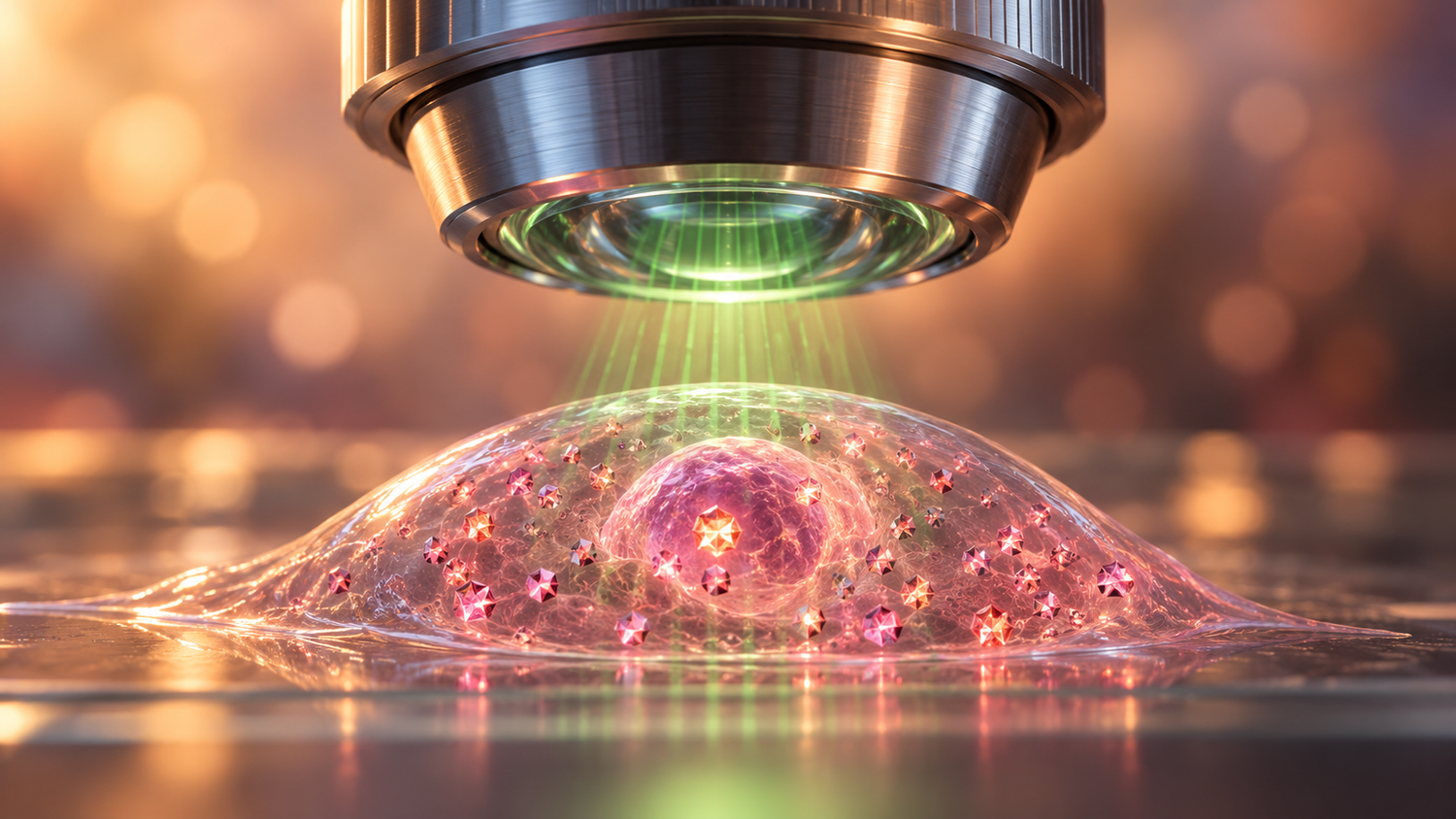
A team at Zhejiang University in Hangzhou has built a microscope that reads diamond quantum sensors inside living human cells at roughly twice the sharpness of conventional fluorescence imaging, and used it to watch reactive oxygen species change from one part of a cell to another. The paper, published in Nature Communications on 10 October 2026, reports that small diamonds in different regions of the same cell responded to two drug treatments in five distinct ways, a variety that a conventional averaged measurement smoothed into a single trend.
The performance figures in the abstract need careful reading, because they come from different experiments. The headline spatial resolution of 136 nanometers was measured on nanodiamond samples outside cells. Inside a live cell the team reports 161 nanometers for the optical image and 178 nanometers for the quantum sensing map. The 150 millisecond figure is the update interval of the sensing images, which the instrument reaches by reusing recent exposures in a rolling reconstruction. The 180 by 180 micrometer field of view is a mosaic of 5 by 5 smaller fields, captured in 12 seconds.
The article in Nature Communications (DOI 10.1038/s41467-026-78321-2) lists Yuxian Lu, Yibo Yang and Ke Xu as equal first authors, with Jiandong Feng as corresponding author. Most of the eleven authors work in the Laboratory of Experimental Physical Biology in Zhejiang University's Department of Chemistry, with co-authors from the university's College of Optical Science and Engineering, Zhejiang Normal University and China Jiliang University. The journal has released the accepted manuscript ahead of the final Version of Record.
How the Zhejiang microscope combines nitrogen-vacancy sensing with structured illumination
The sensing element is the nitrogen-vacancy center, a defect in diamond where a nitrogen atom sits beside a missing carbon atom. Its electron spin responds to temperature and to magnetic noise in its surroundings, and the defect reports that response as a change in red fluorescence. The team fed HeLa cells, a standard human cancer cell line, with fluorescent nanodiamonds about 100 nanometers across, which the cells took up over roughly a day.
The imaging element is structured illumination microscopy, or SIM. The instrument projects fine green stripes onto the sample from a digital micromirror device, records nine images with the stripes rotated and shifted, and computes a picture about twice as sharp as the diffraction limit allows. SIM uses comparatively low light doses, which matters for keeping cells alive during long sessions. The authors call the combined method Super-resolution Multi-physical field Imaging with Deconvolution, or SMID.
Two quantum readouts run on the same setup. One tracks the diamond's spin-relaxation time, known as T1, which shortens when free radicals and other magnetic species are nearby. In calibration tests outside cells, the T1 signal tracked hydroxyl radical concentrations in a range the authors describe as comparable to physiological levels. The other readout follows a shift in the diamond's microwave resonance with temperature, using a fast four-point measurement. A custom three-dimensional locking system holds the sample steady for hour-long recordings.
What the live-cell experiments showed about reactive oxygen species and mitochondria
In live HeLa cells, the team mapped mitochondria, temperature differences and relative reactive oxygen species levels in the same field. Diamonds near mitochondria showed shorter relaxation times, consistent with higher radical levels where cellular respiration takes place. Over an hour of imaging, the mitochondria kept their shape and the diamonds kept a stable brightness, while the mitochondrial dye used for comparison, PK Mito Red, faded progressively.
Quantum pillar: sensing. Technology readiness: TRL 4 of 9. The integrated diamond-sensor microscope has been validated in the laboratory on one human cell line grown in a dish, which places it well before any animal study, patient sample or clinical diagnostic use.
Five response types to superoxide dismutase and CCCP in a single cell
The most interesting result comes from a perturbation experiment. The researchers first added superoxide dismutase, an enzyme that removes superoxide radicals, and then CCCP, a compound that disrupts mitochondrial function and pushes cells toward apoptosis. Measured in the conventional wide-field way, the averaged signal rose after the enzyme and fell after CCCP, a tidy textbook pattern.
Read diamond by diamond, the same cell looked far less tidy. The authors grouped the individual sensors into five response types according to how strongly their signal moved after each treatment, and found that each type showed a preference for particular cell regions. One type showed oscillating radical levels, which the authors suggest may reflect feedback in the cell's antioxidant defenses or rhythmic changes in mitochondrial activity. That interpretation is offered as a possibility, and the study covers a single cell line.
Earlier diamond sensing in mitochondria, and the 2026 thermometry work in Cambridge
Diamond sensors have been used to look for free radicals in cells for several years. In May 2021 a group led by Romana Schirhagl at the University Medical Center Groningen reported in Science Advances that fluorescent nanodiamonds targeted to single mitochondria in macrophage cells could detect radicals generated after stimulation or inhibition, using the same T1 relaxometry. That work followed one targeted sensor at a time. The Zhejiang paper reads many sensors in parallel across a cell, which is what makes the regional comparison possible.
A second line of 2026 work concerns precision. In September a preprint from Cambridge, Warwick and Cardiff described a dual-defect nanodiamond thermometer with an extrapolated resolution floor of 682 microkelvin, an estimate distinct from its measured error of 11.9 millikelvin over 83 seconds. Precise single probes and sharp maps of many probes answer different questions, and instruments that eventually combine both are the likeliest to move into pharmacology laboratories.
What drug developers and hospital research laboratories can take from the result
The nearest users of such an instrument are cell biologists and pharmaceutical teams studying how compounds affect cells. A microscope that maps oxidative stress region by region could help show whether a candidate drug triggers radical bursts in particular parts of a cell, or how mitochondria respond to toxins. Those are preclinical questions, and the paper does not test patient material.
For a hospital research director or a buyer of laboratory equipment, the practical reading is about timing. The setup is custom-built, so a commercial version would need an instrument maker to engineer and validate it. The remaining validation needs are replication in other cell types and laboratories, tests in tissue as well as cultured cells, and independent confirmation that the relaxation signal tracks radical levels quantitatively inside cells. The study was funded by the National Natural Science Foundation of China and Zhejiang provincial programs, the authors declare no competing interests, and the paper is open access.
Sources
Primary source: Yuxian Lu, Yibo Yang, Ke Xu and colleagues, with corresponding author Jiandong Feng (Zhejiang University, Zhejiang Normal University and China Jiliang University), Quantum-sensing-based super-resolution live cell microenvironment imaging, Nature Communications, published 10 October 2026, read in its accepted-manuscript form. Also drawn on: Linyan Nie, Romana Schirhagl and colleagues (University Medical Center Groningen), Science Advances, 21 May 2021, and the Monitor's earlier coverage of nanodiamond thermometry. The readiness rating is the Monitor's editorial assessment.