Does a Quantum Layer Change What a Medical AI Sees?
Quentir Medicine Monitor
Evidence-based insights for quantum medicine. Published by Quentir Systems LLC · July 27, 2026.
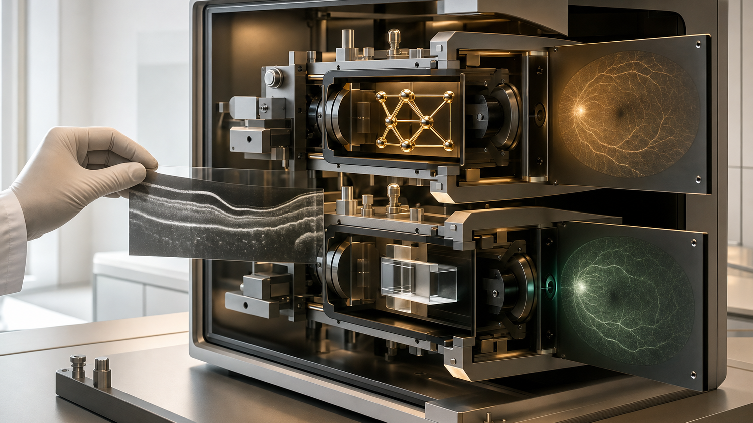
A small, classically emulated quantum circuit changed where a medical-image classifier appeared to concentrate when labeled examples were limited. The advantage did not persist at every sample size: the matched classical network reached the highest retinal accuracy once the training set became large.
A July 2026 preprint makes that comparison unusually clean. Both models used the same convolutional backbone and a comparable number of trainable parameters. They differed in one intermediate layer, creating a parameter-matched comparison between a dense classical layer and a four-qubit circuit computed on ordinary classical hardware. The hybrid model performed best relative to its comparator in an intermediate-data regime, while SHAP attention maps suggested more concentrated retinal focus when data were scarce.
Practical takeaway. The study is a controlled computational benchmark. It reports no physical quantum-processor run, prospective clinical test, clinician reader study, or externally validated diagnostic system.
One substituted layer makes the experiment legible
The central problem in many quantum machine-learning comparisons is attribution. If a hybrid network and its classical comparator have different backbones or very different parameter counts, a performance gap cannot be assigned confidently to the quantum-inspired component. The new preprint by Guillermo Rubiños Rodríguez and colleagues reduces that ambiguity. Each branch begins with the same three-layer convolutional feature extractor. A linear step then compresses the image representation to four values.
In the classical branch, a conventional dense layer transforms those values into a 12-dimensional representation. In the hybrid branch, the four values become rotation angles for four emulated qubits. Trainable rotations and, in one configuration, CNOT gates process the state. Measurements of local Pauli observables produce the same 12-dimensional output expected by the final classifier. The circuit runs in simulation on classical computers, so the test isolates a circuit-shaped representation layer rather than measuring quantum hardware.
The authors trained the pair on retinal optical coherence tomography images from four classes: normal retina, choroidal neovascularization, diabetic macular edema, and drusen. Their search varied training size from 200 to 30,000 images, along with learning rate and batch size. It also tested entanglement and different observable sets. Ten random seeds supported each reported mean in the main retinal comparison. A second task used two-dimensional slices derived from the OASIS-1 structural MRI dataset to sort scans into three classes, from no dementia to a merged mild and moderate group, as a robustness check.
The useful difference appeared between scarcity and abundance
Neither architecture won throughout the retinal experiment. The hybrid model produced its clearest relative gains around 800 and 2,000 training images. At the largest sample sizes, the classical CNN moved ahead and reached the study's highest retinal test accuracy, 93.7 percent. The MRI check followed the same broad pattern: a hybrid advantage at smaller sizes narrowed as more training examples arrived, then reversed slightly in favor of the classical model.
This shape matters more than a single winning score. It suggests that the emulated circuit acted as a particular inductive bias, giving the network a useful way to organize a modest quantity of information. Once the dataset offered enough examples, the classical model reached the better test accuracy. The result therefore concerns where a compact model may be useful, rather than establishing a general hierarchy between quantum-shaped and classical layers.
The retinal source also matters. The image collection originates in the work reported by Daniel Kermany and colleagues in Cell. Reusing a known dataset supports comparison across methods, while it leaves important deployment questions open. A retrospective benchmark cannot show how a model handles a new hospital, a changed scanner, a different patient population, or the disorder mix encountered in routine care.
The explanation maps converged as the dataset grew
Accuracy describes whether a classification was correct. It does not reveal which pixels carried the decision. The researchers therefore compared SHAP attribution maps, which estimate how image regions influence a model's output. Their first tool measured overlap between the top 10 percent of absolute SHAP values from the two models. Their second compared the spatial distributions of positive attributions across the test set.
With 1,000 training images, the hybrid maps appeared more concentrated around retinal layers and disease-related structures. The classical maps were more fragmented in the examples shown. At 30,000 images, overlap increased substantially, indicating that both models had moved toward similar retinal regions. The paper locates the hybrid model's attention in regions affected by the three diseases, particularly around the retinal pigment epithelium and the characteristic elevations produced by the deposits.
That is an interesting interpretability result with a firm limit. An attribution map can show where a model's score is sensitive. It cannot determine whether the highlighted region is a clinically valid biomarker or whether a specialist would use it the same way. The authors explicitly reserve those questions for expert review and external clinical validation. The maps are best read as a consistency test inside this benchmark.
Entanglement did not earn a premium here
The study also asks whether recognizably quantum ingredients were responsible for the hybrid behavior. Removing the CNOT entangling layer produced comparable classification performance and made the circuit much easier to simulate as qubit count increased. In this four-qubit setting, the simpler circuit carried much of the useful behavior.
Measurement design showed a separate dependency. Larger observable sets could describe pairwise or three-qubit correlations, increasing the feature space available to the classifier. They helped only when the training set was sufficiently large. With limited data, the smaller local-observable set performed comparably while demanding less measurement work in a hypothetical hardware implementation.
These findings narrow the interpretation. The hybrid layer may provide a useful representational constraint under selected data conditions. The benchmark does not show that entanglement created the gain. Its classically simulated circuit avoids processor noise and finite-shot uncertainty, along with decoherence and hardware-connectivity constraints. A later hardware experiment would be a different test with a different cost structure.
The medical record stops well before deployment
The OCT task used labeled images from an established research dataset. The dementia task reduced three-dimensional MRI volumes to selected two-dimensional slices, which limits anatomical context. The paper reports no external validation on images collected at another institution. It includes no prospective workflow, no comparison with clinician readers, and no measurement of patient outcomes.
Those omissions are appropriate for an architecture benchmark, yet they determine what the result can mean for medicine. Before a model could support care, investigators would need to test calibration as well as accuracy, examine performance across clinically relevant groups, and evaluate robustness to acquisition changes. Specialists would need to judge whether the highlighted anatomy is useful and whether the system changes decisions in a beneficial way.
There is also a governance lesson in the study's design. A model claim becomes more informative when the comparator differs by one declared component and the training regime is varied openly. That discipline exposes conditional behavior that a headline accuracy could hide. It shows when the hybrid layer helped, when the classical layer recovered, and which quantum-style features turned out to be unnecessary.
How Quentir Reads It
Quentir reads this preprint as a well-controlled account of conditional model behavior. The result is neither a clinical quantum advantage nor an empty simulation. A four-qubit circuit, calculated classically, changed the intermediate representation enough to affect accuracy and attribution patterns under selected sample sizes. More data reduced that distinction and ultimately favored the matched classical network.
The next useful record would combine an independently collected imaging cohort with specialist assessment of the attribution maps. It would also need a prospective comparison that measures decisions rather than labels alone. A hardware run would answer a separate engineering question about noise and resource use across measurement and runtime. Keeping those steps distinct protects an intriguing computational result from being mistaken for medical readiness.
The enduring question is precise. When labeled retinal images are limited, does a compact circuit-shaped layer change how an equally sized network performs and where it looks? This paper answers with two measured differences at selected OCT sample sizes: higher accuracy in the intermediate-data regime, and attribution maps concentrated on disease-related retinal regions. Its separate OASIS robustness check adds directional support for the performance trend, without extending the retinal SHAP finding to brain anatomy.
Sources
Primary source: Rubiños Rodríguez, Ottavianelli, Alonso, Blázquez Gil, Rauchmann, Díez-Valle, and Altares-López, arXiv preprint submitted July 23, 2026. Dataset context: Kermany and colleagues, Cell, February 22, 2018; and the OASIS Brains project.